Iron deficiency anemia
Board exam relevance: in 13 of 105 exam reports · rank 17- Synonyms
- iron deficiency, low iron, sideropenic anaemia, low ferritin
- Specialty
- Internal medicine · Haematology & oncology
- Images
- Clinical 2 · Blood smear & cytology 2
- Last updated
- 10/2026 · Dr. Pascal Bafteh
Contents
Images (4)
 Blood smear & cytology
 Blood smear & cytology
Definition
Iron deficiency is a reduction in total body iron. Iron deficiency anemia is present when hemoglobin falls below the age- and sex-specific normal value as a result of iron deficiency (according to the WHO 12 g/dl in women, 13 g/dl in men).
It is the classic hypochromic, microcytic anemia and the most common form of anemia overall. Because it usually results from blood loss, finding iron deficiency always prompts a search for the source of bleeding.
Classification
Stages
- Stage I – storage iron deficiency: iron stores are reduced (low ferritin), erythropoiesis is still adequately supplied; hemoglobin and blood count are normal.
- Stage II – iron-deficient erythropoiesis: red cell precursors are no longer adequately supplied with iron; transferrin saturation falls, soluble transferrin receptor and zinc protoporphyrin rise, hemoglobin is still within the normal range.
- Stage III – iron deficiency anemia: hemoglobin below the normal value, with increasing microcytosis and hypochromia.
The terms “latent” and “prelatent” iron deficiency are considered outdated. Functional iron deficiency is distinguished from absolute iron deficiency: stores are replete, but the iron is unavailable to erythropoiesis – typical of anemia of chronic disease.
Occurrence & epidemiology
Iron deficiency is the most common nutritional deficiency worldwide and, at no less than 50 %, the most common cause of anemia; an estimated two billion people are affected. In Europe its prevalence is 5–10 %, in women of reproductive age about 20 %. Other risk groups are infants and young children, especially between 6 and 24 months of age, adolescents during growth (4–8 % at age 13–15, mostly as storage iron deficiency) and pregnant women, 20–30 % of whom develop iron deficiency anemia.
Aetiopathogenesis
Iron metabolism
The body contains about 3–5 g of iron, roughly 3 g of it in hemoglobin. Stores amount to about 0.5–1 g in men but only 0.2–0.4 g in premenopausal women. Body iron is regulated exclusively through absorption, as active excretion is not possible.
Iron is absorbed in the duodenum and upper jejunum. Hem iron from meat is absorbed much better than ferric non-hem iron from plant foods, which is first reduced to ferrous iron. The central regulator is hepcidin, produced in the liver: it causes degradation of the iron exporter ferroportin and thus throttles iron uptake from the gut and release from macrophages. In iron deficiency hepcidin production falls and absorption rises.
Causes
- Blood loss (most common cause): in men and postmenopausal women mostly chronic occult bleeding from the gastrointestinal tract (ulcer, carcinoma, polyps, angiodysplasia, hemorrhoids, reflux esophagitis); in premenopausal women mainly menstruation – about 15 % of women have heavy menstrual bleeding with more than 80 ml of blood loss per cycle; also blood donation, urinary or pulmonary blood loss, and worldwide frequently hookworm infection.
- Increased requirements: growth in infancy, early childhood and adolescence, pregnancy and lactation, high-performance sport, chronic intravascular hemolysis.
- Decreased absorption: unbalanced or strict vegetarian diet, anorexia, atrophic or Helicobacter pylori gastritis, achlorhydria, celiac disease, inflammatory bowel disease, short bowel syndrome.
- Genetic (rare): iron-refractory iron deficiency anemia (IRIDA) due to autosomal recessive mutations in the TMPRSS6 gene (matriptase-2) with inappropriately high hepcidin.
Clinical features
Symptoms and signs
Symptoms combine general signs of anemia with the specific consequences of iron deficiency. Their severity depends on the degree of anemia and the speed at which it develops.
- fatigue, reduced performance, exertional dyspnea, dizziness, tachycardia, conjunctival pallor
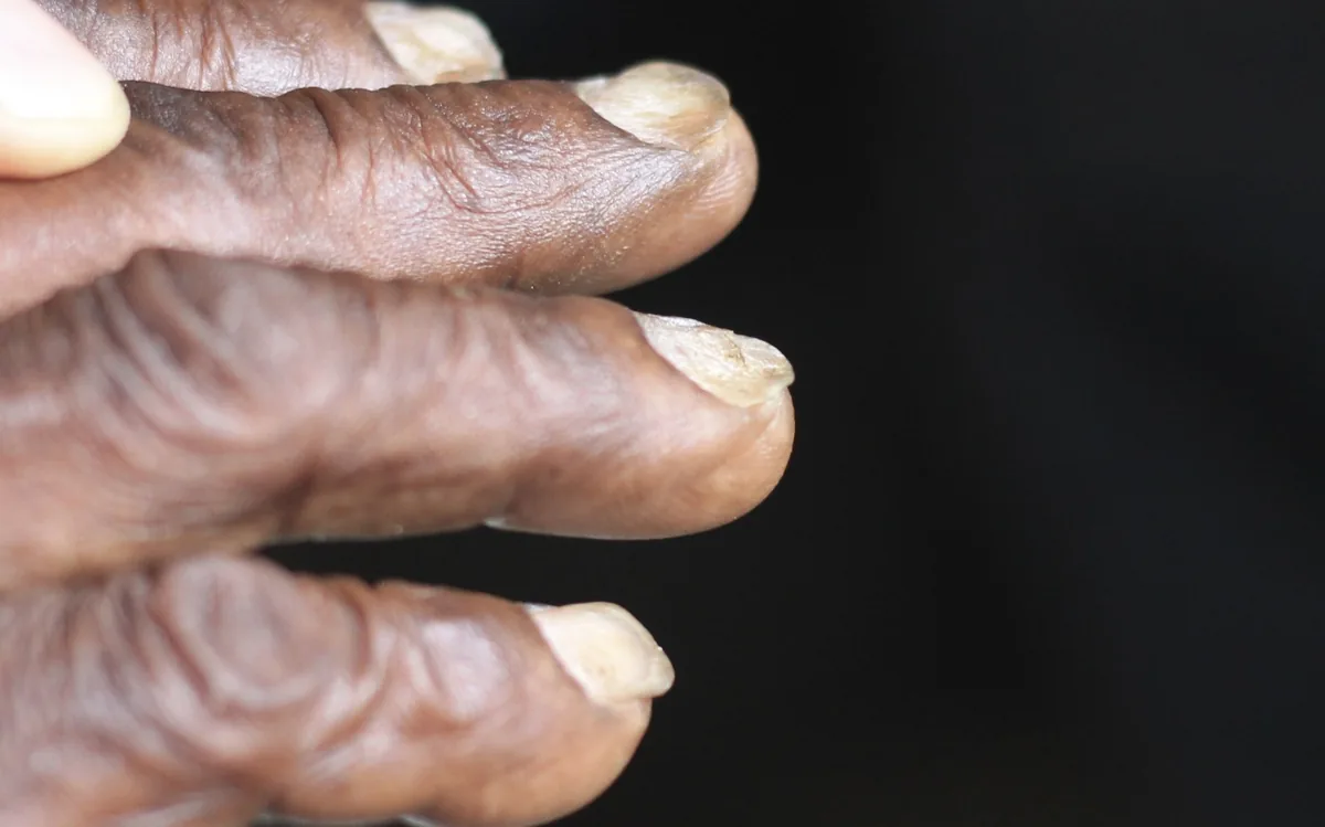
- brittle nails and hair, spoon-shaped nails (koilonychia)
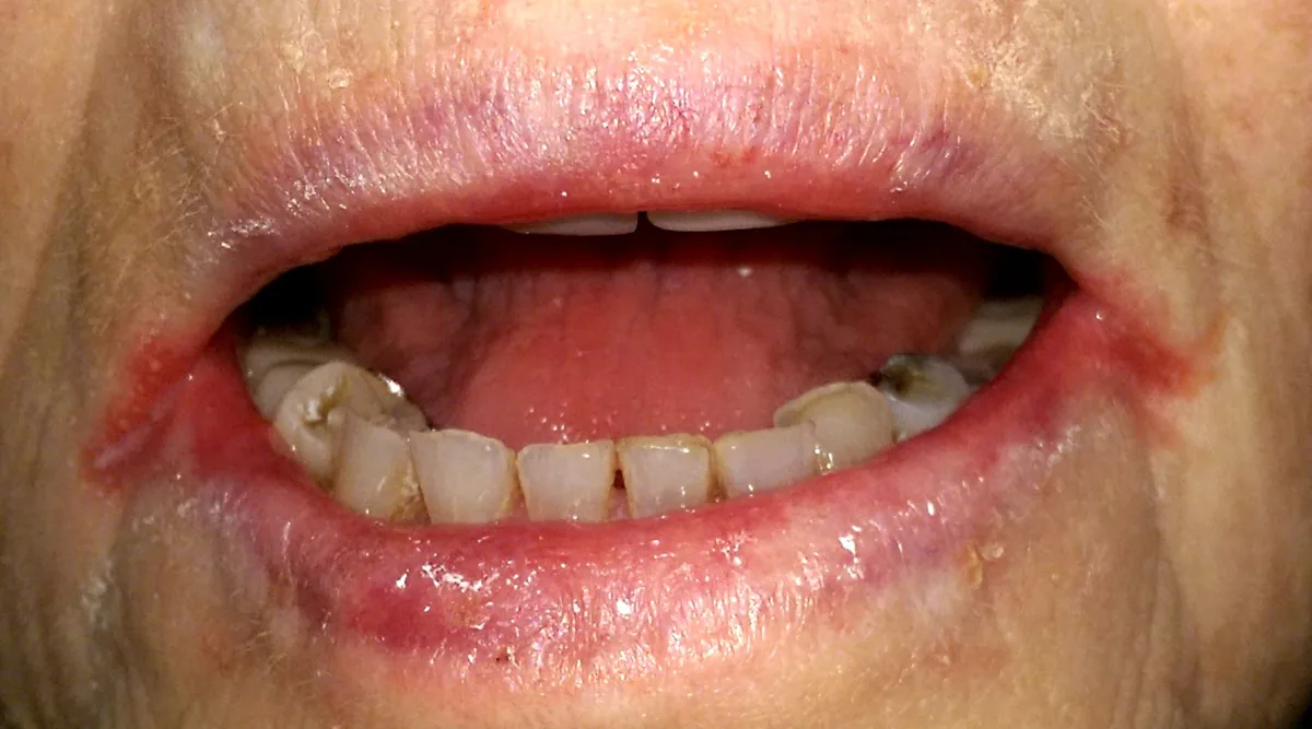
- angular cheilitis, atrophic glossitis, dysphagia
- pica: craving for non-food substances such as ice, earth or starch
- restless legs syndrome
- impaired concentration, headache, sleep disturbance, depressed mood
- in children with severe chronic iron deficiency growth disturbance as well as neurological and cognitive deficits
- in pregnancy with severe anemia more frequent miscarriage, preterm birth and fetal developmental disorders
Histology
Blood smear and bone marrow
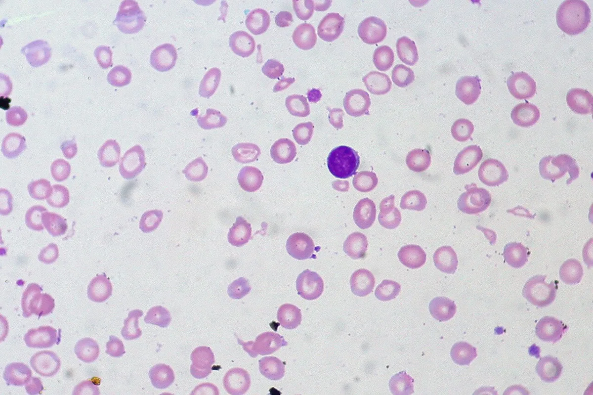
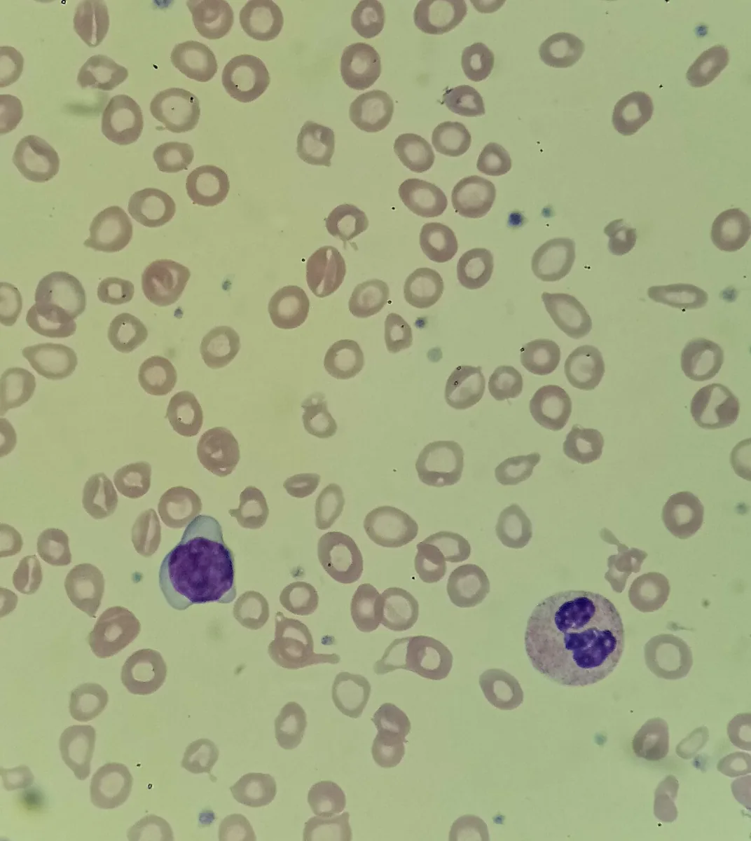
- Smear: hypochromic, microcytic red cells with enlarged central pallor, ring-shaped cells (anulocytes), cigar-shaped cells and elliptocytes, marked anisocytosis and poikilocytosis.
- Platelets: reactively increased in 20–30 %.
- Bone marrow (Prussian blue stain): absent storage iron; sideroblasts make up less than 15 % of red cell precursors (normal 15–50 %). Bone marrow examination is the gold standard but is performed for this question only in exceptional cases.
Diagnosis
Blood count
- MCV below 80 fl and MCH below 28 pg (hypochromic, microcytic); in the early stage the anemia may still be normocytic.
- increased red cell distribution width (RDW) reflecting anisocytosis
- reticulocytes normal or low, raised with ongoing bleeding
- Reticulocyte hemoglobin content (CHr): an early marker; values below about 26 pg indicate iron-deficient erythropoiesis.
- Hypochromic red cells (HYPO): a proportion above 10 % is regarded as proof of iron-deficient erythropoiesis (normal below 2.5 %).
Iron parameters
- Ferritin: the most sensitive marker; it correlates with iron stores and falls as early as stage I. Values below 15 µg/l are regarded as diagnostic; increasingly, below 30 µg/l regardless of sex is used as the cut-off in adults. As an acute-phase protein, ferritin rises in inflammation, malignancy and liver disease and can mask iron deficiency; with inflammation, values up to about 100 µg/l are still compatible with iron deficiency.
- Transferrin saturation: normal 16–45 %; values of 15 % or less indicate iron-deficient erythropoiesis. It is also low in inflammation and varies during the day.
- Transferrin or total iron-binding capacity: raised.
- Soluble transferrin receptor: rises from stage II; usually normal in functional iron deficiency due to inflammation.
- Serum iron: unsuitable for diagnosis because of diurnal variation and because it is also low in inflammation.
Search for the cause
Once the diagnosis is made, occult bleeding is assumed to be the cause until proven otherwise. In men and postmenopausal women without an obvious source of bleeding, examination of the gastrointestinal tract (gastroscopy, colonoscopy) is part of the work-up, because anemia may be the only sign of an occult carcinoma. Depending on the history, tests for celiac disease and Helicobacter pylori, urinalysis for blood and questions about blood donation, menstrual flow, nosebleeds and diet are added.
Keep learning in the app
Further reading (open access)
Cross-references
More topics: Haematology & oncology
Note: Learning content for medical education – not a treatment recommendation and no substitute for diagnosis or treatment decisions in individual cases. Treatment and management are deliberately not covered on this page.