Hemolytic anemias
Board exam relevance: in 14 of 105 exam reports · rank 14- Synonyms
- haemolysis, hemolytic anemia, red cell destruction, intravascular haemolysis, extravascular haemolysis
- Specialty
- Internal medicine · Haematology & oncology
- Images
- Blood smear & cytology 3
- Last updated
- 10/2026 · Dr. Pascal Bafteh
Contents
Images (3)
 Blood smear & cytology
 Blood smear & cytology
 Blood smear & cytology
Definition
Hemolysis is the premature destruction of red blood cells, with a lifespan shorter than the normal approximately 120 days. Hemolytic anemia develops when the bone marrow can no longer make up for the shortened red cell lifespan (uncompensated hemolysis). If the marrow compensates by increased production, the condition is called compensated hemolysis without anemia.
Classification
By cause
Corpuscular (intrinsic) causes – a defect of the red cell itself, usually inherited:
- membrane defects: hereditary spherocytosis, elliptocytosis, stomatocytosis; acquired: paroxysmal nocturnal hemoglobinuria, severe hypophosphatemia, acanthocytosis in liver disease
- enzyme defects: G6PD deficiency (hexose monophosphate shunt), pyruvate kinase deficiency (glycolysis)
- hemoglobinopathies: sickle cell disease, thalassaemias, HbC and HbE disease, unstable hemoglobins
Extracorpuscular (extrinsic) causes – external factors, usually acquired:
- immunological: autoimmune hemolysis (warm and cold antibodies, paroxysmal cold hemoglobinuria), drug-induced immune hemolysis, alloantibody-mediated hemolysis in blood group incompatibility
- mechanical: thrombotic microangiopathies (TTP, HUS), DIC, HELLP syndrome, defective or mechanical heart valves, march hemoglobinuria
- infectious: malaria, babesiosis, Bartonella; Clostridium perfringens toxins; Shiga toxin
- toxic and physical: oxidising substances (e.g. naphthalene), lead, copper (Wilson disease), snake and insect venoms, burns
- hypersplenism
By site and course
- Extravascular hemolysis: breakdown of damaged or antibody-coated red cells by macrophages in the spleen and liver; the more common form. Spherocytes on the smear and splenomegaly are typical.
- Intravascular hemolysis: destruction of cells directly within the circulation by complement, shear stress, trauma or toxins; leads to free hemoglobin in plasma, hemoglobinuria and hemosiderinuria.
- Course: acute (hemolytic crisis), chronic or episodic.
Aetiopathogenesis
Pathophysiology
Senescent red cells are normally removed by phagocytes in the spleen, liver and bone marrow. The iron is recycled, while hem is degraded to bilirubin, glucuronidated in the liver and excreted in bile.
Extravascular: the spleen removes even mildly altered cells or cells coated with warm antibodies; an enlarged spleen retains even normal red cells. Severely damaged cells or cells coated with IgG or complement (C3) are additionally cleared in the liver with its large blood flow.
Intravascular: when released hemoglobin exceeds the binding capacity of haptoglobin, hemoglobinaemia results and haptoglobin falls. Free hemoglobin dimers are filtered by the glomeruli and reabsorbed by the tubules; once this capacity is exhausted, hemoglobinuria appears. Iron is stored as hemosiderin in tubular cells and reaches the urine when these cells slough.
Consequences: when bilirubin production exceeds the conjugating capacity of the liver, indirect hyperbilirubinaemia and jaundice result; more stercobilin is excreted in stool and more urobilinogen in urine, and gallstones can form. Increased erythropoietin production in response to anemia leads to reticulocytosis.
Clinical features
- general signs of anemia: pallor, fatigue, dizziness, weakness
- scleral icterus or jaundice without liver disease
- splenomegaly
- dark, reddish-brown urine with hemoglobinuria
- gallstones in chronic hemolysis
- increased tendency to thrombosis in many forms of hemolysis
- Hemolytic crisis (acute, severe hemolysis, uncommon): chills, fever, back and abdominal pain, shock
Histology
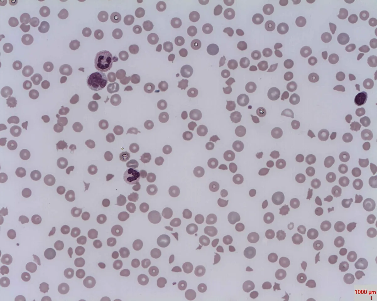
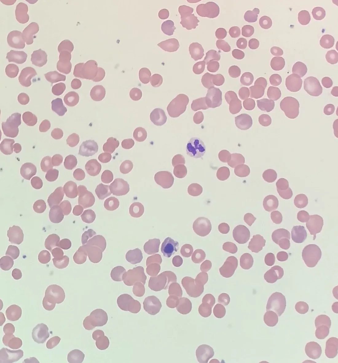
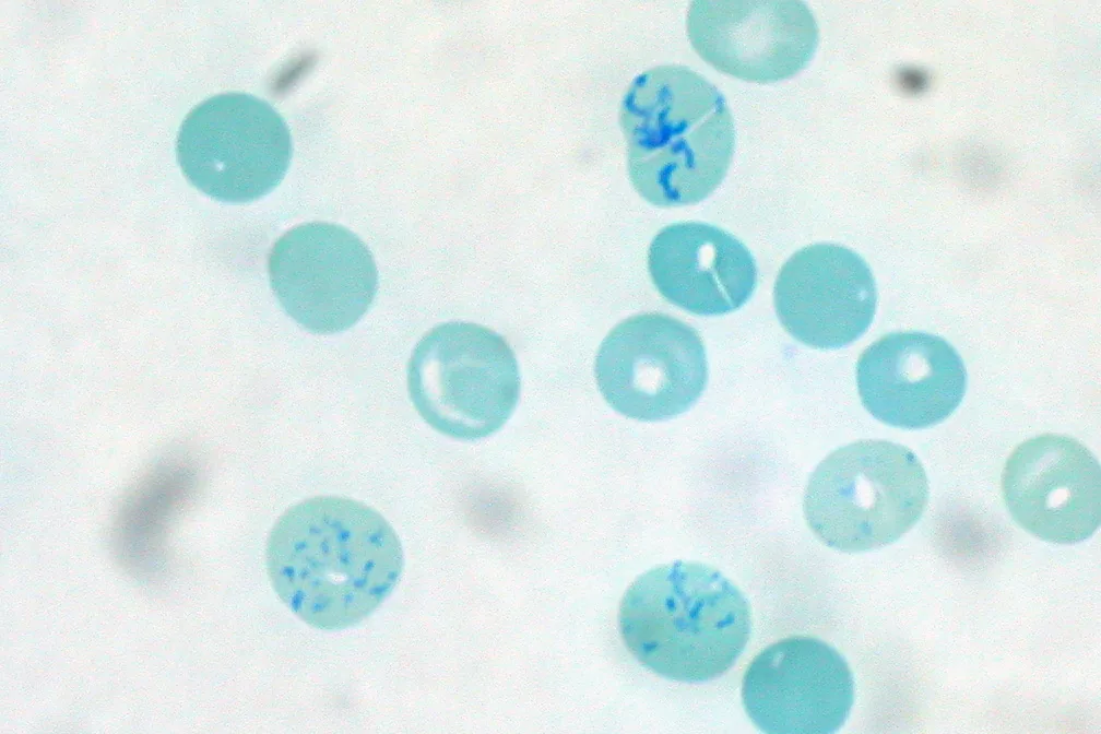
Blood smear
The blood smear and reticulocyte count are the most important tests for detecting hemolysis; red cell morphology often points to the cause.
- Spherocytes: extravascular hemolysis – hereditary spherocytosis, warm autoimmune hemolysis
- Schistocytes (fragmented cells): intravascular mechanical hemolysis – TTP, HUS, DIC, valvular hemolysis; also in severe B12 deficiency (pseudo-TTP)
- Agglutinates: cold agglutinin disease (if the sample is not kept warm)
- Heinz bodies, bite and blister cells: oxidative stress, G6PD deficiency, unstable hemoglobins
- Sickle cells: sickle cell disease
- Codocytes (bullseye cells): hemoglobinopathies, liver disease, after splenectomy
- Nucleated red cell precursors and basophilic stippling: thalassaemia major
- Acanthocytes and echinocytes: liver and kidney disease, glycolytic defects
- Polychromasia: reflects reticulocytosis
Diagnosis
Confirming hemolysis
- Reticulocytes raised. If the rise fails to occur (e.g. in renal insufficiency, infection or marrow failure), rapidly severe anemia may follow.
- LDH and indirect bilirubin raised, haptoglobin low. Haptoglobin can fall in hepatic dysfunction without hemolysis and may be normal despite hemolysis in inflammation.
- Urine: urobilinogen raised; in intravascular hemolysis hemoglobinuria (dipstick positive for blood but no red cells on microscopy) and hemosiderinuria.
- Plasma: reddish-brown due to free hemoglobin in intravascular hemolysis, in contrast to myoglobinaemia.
- Full blood count: accompanying thrombocytopenia raises suspicion of thrombotic microangiopathy, DIC or PNH.
Establishing the cause
- History: ethnic origin, family history, underlying disorders, drug and toxin exposure, infections, travel
- Examination for splenomegaly
- Direct antiglobulin test (direct Coombs test): detects IgG or complement (C3) bound to red cells; positive in autoimmune hemolysis. After receipt of donor blood within the last 3 months, a positive result may also reflect alloantibodies.
- Indirect Coombs test: free antibodies against red cells in serum.
- Hemoglobin analysis (electrophoresis, HPLC), enzyme assays (e.g. G6PD, pyruvate kinase), flow cytometry for a PNH clone, cold agglutinins, osmotic fragility or membrane tests, genetic testing
- When thrombotic microangiopathy is suspected: ADAMTS13 activity, Shiga toxin testing if diarrhea is present, complement studies where appropriate
Keep learning in the app
Further reading (open access)
Cross-references
More topics: Haematology & oncology
Note: Learning content for medical education – not a treatment recommendation and no substitute for diagnosis or treatment decisions in individual cases. Treatment and management are deliberately not covered on this page.