Anemia (classification and work-up)
Board exam relevance: in 33 of 105 exam reports · rank 1- Synonyms
- low blood count, low haemoglobin, low hemoglobin, anemia, microcytic anaemia, macrocytic anaemia
- Specialty
- Internal medicine · Haematology & oncology
- Images
- Clinical 1 · Blood smear & cytology 2
- Last updated
- 10/2026 · Dr. Pascal Bafteh
Contents
Images (3)
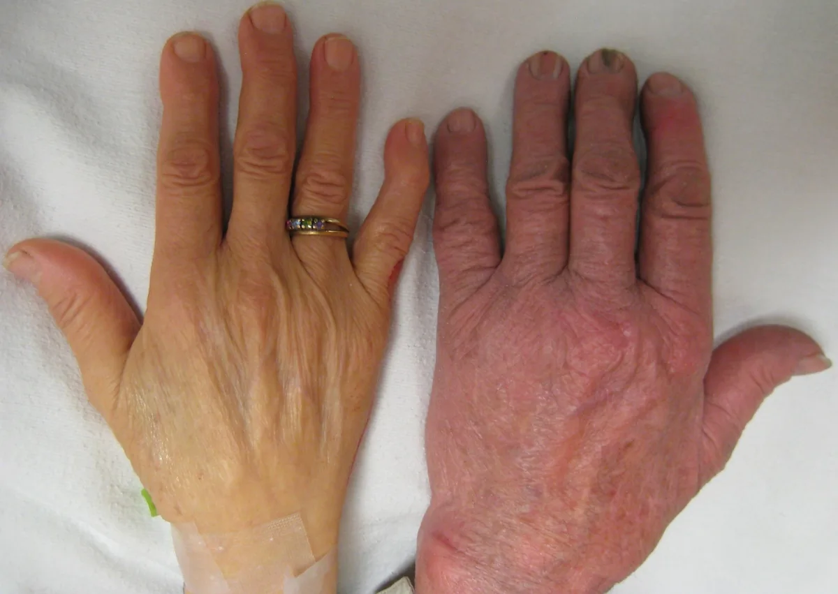
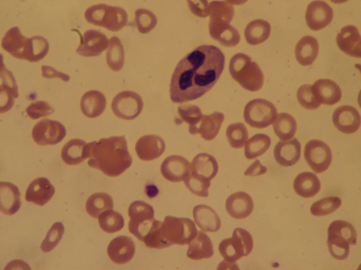 Blood smear & cytology
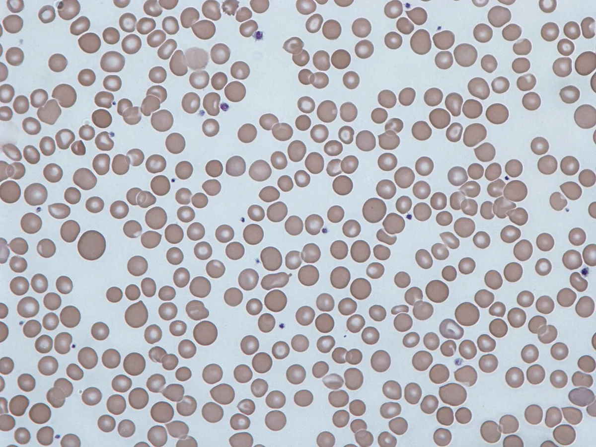 Blood smear & cytology
Definition
Anemia is a reduction in red blood cells, measured as hemoglobin (Hb), hematocrit or red cell count. According to the WHO it is present in adults when Hb falls below 13 g/dl (men) or below 12 g/dl (women). In pregnancy, lower limits are used because of the expanded plasma volume (lower reference value 11.0 g/dl in the first and third trimester, 10.5 g/dl in the second).
Anemia is not a diagnosis in itself but a manifestation of an underlying disorder. Even a mild, asymptomatic anemia therefore always has a cause that needs to be identified.
Classification
By red cell size (MCV)
- Microcytic (MCV below 80 fl): impaired hem or globin synthesis – iron deficiency, thalassaemia, anemia of chronic disease (in part), sideroblastic anemia, iron transport defect.
- Normocytic (MCV 80–100 fl): anemia of chronic disease, kidney disease, endocrine failure (thyroid, pituitary), myelodysplasia, marrow infiltration (myelophthisis), parvovirus B19 infection, pure red cell aplasia, acute hemorrhage before iron deficiency develops.
- Macrocytic (MCV above 100 fl): impaired DNA synthesis in vitamin B12 or folate deficiency, also alcohol use disorder, liver disease, myelodysplasia, copper deficiency; marked reticulocytosis can also raise the MCV.
Occurrence & epidemiology
Anemia is one of the most common health problems worldwide and mainly affects young children, pregnant women and women of reproductive age. According to WHO estimates, about 40 % of children aged 6–59 months, 37 % of pregnant women and 30 % of women aged 15 to 49 years have anemia. The most common cause is iron deficiency, which accounts for at least half of all anemias; regionally, hemoglobinopathies and malaria play a major role.
Aetiopathogenesis
Red cell mass reflects the balance between production on the one hand and destruction or loss on the other. Anemia therefore results from blood loss, from insufficient production or from increased destruction – often from a combination.
Acute blood loss: the fall in Hb often becomes visible only after several hours, when interstitial fluid moves into the vascular space and dilutes the remaining red cells. During the first hours granulocytes and platelets rise.
Chronic blood loss causes anemia when losses exceed new production or – more commonly – when iron stores are exhausted.
Impaired production: if erythropoiesis stops completely, the red cell count falls by about 7–10 % per week (roughly 1 % per day).
Hemolysis: antibodies or complement on the cell surface, inherited defects or an enlarged spleen shorten red cell lifespan. Reticulocyte production normally rises in response, as long as iron, vitamins and erythropoietin are sufficient.
Clinical features
General signs of anemia
Symptoms result from tissue hypoxia and its compensation; they are neither sensitive nor specific and do not distinguish between types of anemia. They are more pronounced when anemia develops rapidly or heart or lung disease is present.
- fatigue, weakness, reduced exercise tolerance, exertional dyspnea
- pallor of skin and mucous membranes, especially in severe anemia (Hb below 7 g/dl)
- dizziness, headache, pulsatile tinnitus, drowsiness
- tachycardia, angina pectoris, syncope
- amenorrhoea, loss of libido, gastrointestinal complaints
- in severe hypoxia or hypovolaemia heart failure (rarely high-output heart failure) or shock
Clues to the cause
- melaena, hematochezia, hematemesis, epistaxis or heavy menstruation: bleeding
- jaundice and dark urine without liver disease: hemolysis
- weight loss: malignancy
- diffuse, severe bone or chest pain: sickle cell disease
- stocking-glove paraesthesias, peripheral neuropathy: vitamin B12 deficiency
- splenomegaly: hemolysis, hemoglobinopathy, myeloproliferative disorder, infection, lymphoma
- fever and heart murmur: infective endocarditis
Histology
Blood smear
- Schistocytes (fragmented cells): mechanical damage in microangiopathic hemolytic anemia (TTP, HUS, DIC) or prosthetic heart valves
- Spherocytes: hereditary spherocytosis, warm autoimmune hemolysis
- Bite and blister cells, Heinz bodies: oxidative damage, e.g. in G6PD deficiency
- Codocytes (bullseye cells): thalassaemia, other hemoglobinopathies, liver disease, after splenectomy
- Sickle cells: sickle cell disease
- Teardrop cells and nucleated precursors: marrow infiltration or myelofibrosis
- Hypersegmented neutrophils and oval macrocytes: megaloblastic anemia
Diagnosis
Basic laboratory tests
- Full blood count with white cells and platelets: abnormalities of several cell lines raise suspicion of a primary bone marrow disorder.
- Red cell indices: MCV (cell volume), MCH (Hb content per cell), MCHC (Hb concentration; raised with spherocytes) and RDW (size distribution). A raised RDW with a normal MCV may be the only sign of coexisting microcytic and macrocytic processes.
- Reticulocytes (relative, absolute or as reticulocyte production index): reflect the marrow response. Raised values point to hemolysis, bleeding or recovery; low or normal values in anemia indicate inadequate production. They can be low even in hemolysis, for example with coexisting kidney disease or infection.
- Blood smear: cell morphology, confirmation of thrombocytopenia (exclusion of pseudothrombocytopenia due to platelet clumping), parasites such as plasmodia.
Further investigations
- Iron status: ferritin, transferrin saturation, soluble transferrin receptor where available
- Vitamin B12 and folate, with borderline results methylmalonic acid and homocysteine
- Hemolysis markers: LDH and indirect bilirubin raised, haptoglobin low; in bleeding LDH and bilirubin are normal.
- Direct antiglobulin test (Coombs test) when immune hemolysis is suspected
- Hemoglobin analysis (e.g. electrophoresis, HbA2, HbF) when a hemoglobinopathy is suspected
- Creatinine, inflammatory markers, thyroid tests, urinalysis and fecal occult blood test depending on the suspected cause
- Bone marrow examination: in unexplained anemia, with abnormalities of more than one cell line or suspected primary marrow disease (leukemia, myeloma, aplastic anemia, myelodysplasia, myelofibrosis, metastatic carcinoma)
Keep learning in the app
Further reading (open access)
Cross-references
More topics: Haematology & oncology
Note: Learning content for medical education – not a treatment recommendation and no substitute for diagnosis or treatment decisions in individual cases. Treatment and management are deliberately not covered on this page.