Thalassemia
Board exam relevance: in 2 of 105 exam reports · rank 142- Synonyms
- Mediterranean anaemia, Cooley anaemia, beta thalassaemia, alpha thalassaemia, thalassaemia trait
- Specialty
- Internal medicine · Haematology & oncology
- Images
- Blood smear & cytology 2
- Last updated
- 10/2026 · Dr. Pascal Bafteh
Contents
Images (2)
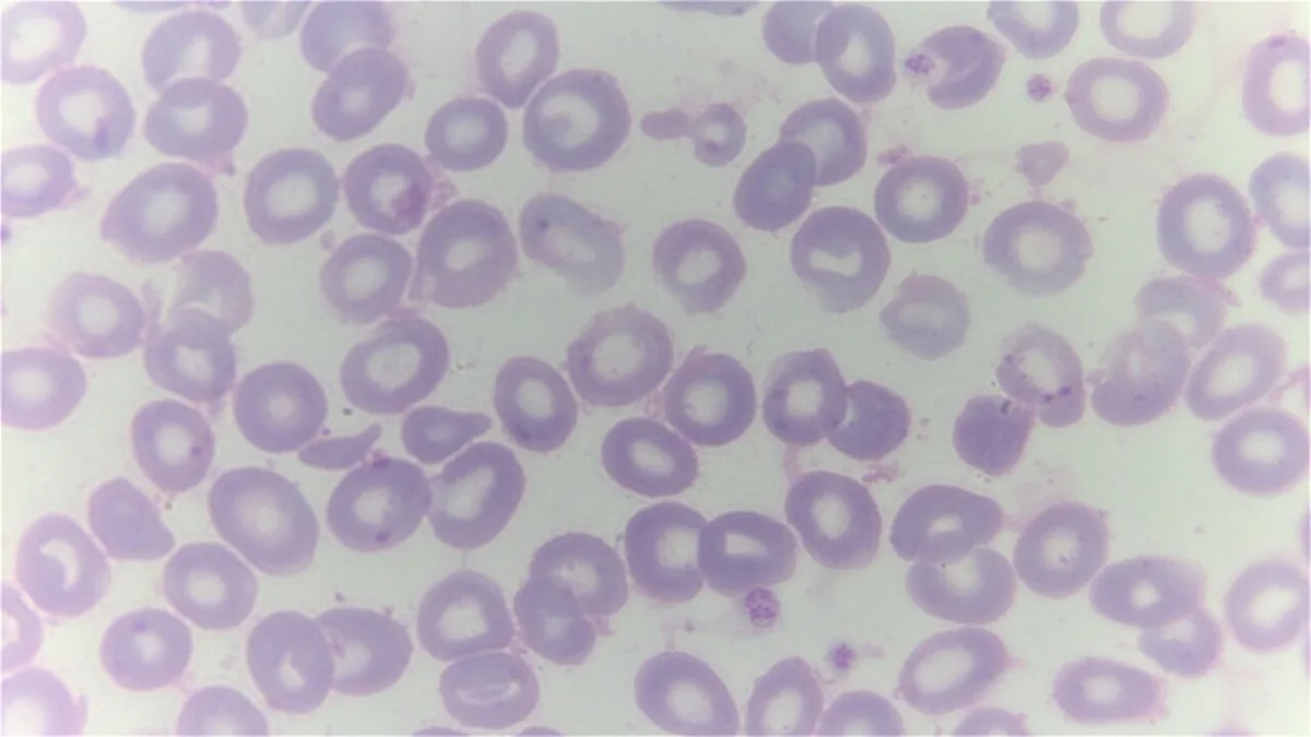 Blood smear & cytology
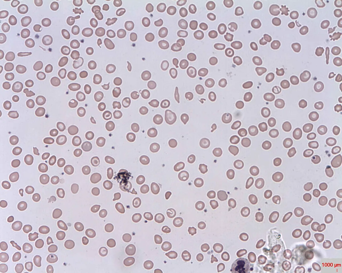 Blood smear & cytology
Definition
Thalassaemias are a group of mostly autosomal recessive, microcytic anemias in which the synthesis of one or more globin chains is reduced or absent. Depending on the affected gene they are called α- or β-thalassaemia. They belong to the hemoglobinopathies and are among the most common monogenic hereditary disorders worldwide.
Normal adult hemoglobin (HbA) consists of two α and two β chains; in addition there is up to 2.5 % HbA2 (α2δ2) and less than 1.4 % fetal hemoglobin HbF (α2γ2).
Classification
α-thalassaemia (four α-globin genes on chromosome 16)
- One gene affected: silent carriers, clinically normal
- Two genes affected (α-thalassaemia minor): mild to moderate microcytic anemia without symptoms
- Three genes affected (HbH disease): excess β chains form tetramers (HbH), in infants γ tetramers (Hb Bart's); symptomatic hemolytic anemia with splenomegaly
- Four genes affected: hydrops fetalis, not viable in utero, because hemoglobin lacking α chains cannot transport oxygen
β-thalassaemia (β-globin gene on chromosome 11)
Depending on residual gene activity, β⁰ alleles (no production) and β⁺ alleles (reduced production) are distinguished. Clinically, three basic types are recognized:
- β-thalassaemia minor (heterozygous carriers): usually asymptomatic, mild microcytic anemia
- β-thalassaemia intermedia (mostly homozygous or compound heterozygous): broad spectrum, Hb often around 7–10 g/dl, presentation from childhood to adulthood
- β-thalassaemia major (Cooley anemia; homozygous β⁰/β⁰ or severe compound heterozygous): severe anemia within the first two years of life
Occurrence & epidemiology
α-thalassaemia is particularly common in people of African, Mediterranean or Southeast Asian origin, β-thalassaemia in the Mediterranean region, the Middle East, the Indian subcontinent, Southeast Asia and Africa. There the proportion of carriers is 5–30 %. In the population of German descent, heterozygous β-thalassaemia is very rare at an estimated 0.01 %; through migration, however, the number of hemoglobinopathy carriers in Germany has risen considerably (more than 400,000 as early as 2010).
Aetiopathogenesis
α-thalassaemia is mostly due to deletions of α-globin genes, β-thalassaemia predominantly to point mutations in the HBB gene (more than 260 variants known). The key factor is globin chain imbalance: in β-thalassaemia the excess of α chains damages the red cell precursors. The result is highly ineffective erythropoiesis with massive marrow expansion, milder hemolysis and hypochromic anemia. The increased but ineffective erythropoiesis suppresses hepcidin and thus increases intestinal iron absorption.
Severity is modulated by residual gene activity and by modifying factors, particularly coexisting α-thalassaemia or hereditary persistence of HbF. Related variants with a thalassaemic picture are the HbE hemoglobinopathy, common in Southeast Asia, and Hb Lepore.
Clinical features
Minor forms are usually asymptomatic or only mildly symptomatic. Coexisting iron deficiency, for example in pregnancy or in children, can markedly worsen the anemia.
Thalassaemia major and severe intermedia:
- severe anemia with pallor, impaired growth, delayed or absent puberty
- jaundice, gallstones, leg ulcers
- pronounced, often massive splenomegaly, hypersplenism
- marrow expansion: thickening of the skull bones and malar eminences, pathological fractures of the long bones
- extramedullary hematopoiesis, typically paravertebral, in more than 20 % of thalassaemia intermedia
- iron overload with cardiomyopathy and heart failure, hepatic siderosis progressing to cirrhosis and endocrine disorders (hypogonadism in up to two thirds, diabetes mellitus in up to 15 %)
Histology
Blood smear and bone marrow
Small, pale red cells, codocytes (bullseye cells) and basophilic stippling are typical; in thalassaemia major the smear with numerous nucleated red cell precursors is virtually diagnostic. The bone marrow shows marked erythroid hyperplasia.
Diagnosis
- Blood count: microcytosis with a red cell count that is high relative to Hb; in carriers often an incidental finding. In thalassaemia major Hb is frequently 6 g/dl or less.
- Hemolysis and iron parameters: bilirubin, serum iron and ferritin raised.
- Quantitative hemoglobin analysis (electrophoresis, HPLC): β-thalassaemia minor with mildly raised HbA2 (above 3.5–4 %); in thalassaemia major HbF markedly raised (up to 90 %) and HbA2 above 3 %. In α-thalassaemia HbA2 and HbF are usually normal; HbH disease shows fast-migrating HbH or Hb Bart's fractions.
- Molecular genetics: confirmation particularly of α-thalassaemia, in unclear β findings and as the basis for prenatal diagnosis.
- X-ray: in thalassaemia major thinned cortex, widened diploic space with a radiating (“sun-ray”) trabecular pattern, osteoporosis of long bones, paravertebral foci of hematopoiesis on chest imaging.
Keep learning in the app
Further reading (open access)
Cross-references
More topics: Haematology & oncology
- Anemia (classification and work-up)
- Hemolytic anemias
- Acute myeloid leukemia (AML)
- Iron deficiency anemia
- Non-Hodgkin lymphomas
- Vitamin B12 deficiency and pernicious anemia
- Multiple myeloma
- Paraneoplastic syndromes
- Autoimmune hemolytic anemia (AIHA)
- Febrile neutropenia
- Immune thrombocytopenia (ITP)
- Renal anemia
Note: Learning content for medical education – not a treatment recommendation and no substitute for diagnosis or treatment decisions in individual cases. Treatment and management are deliberately not covered on this page.