Superior vena cava syndrome
- Synonyms
- SVC syndrome, SVCS, superior vena cava obstruction, SVC obstruction
- Specialty
- Internal medicine · Haematology & oncology
- Images
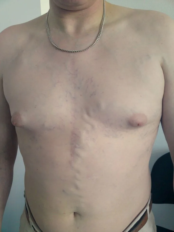
- Clinical 1 · CT 1 · X-ray 1 · Gross specimen 1
- Last updated
- 10/2026 · Dr. Pascal Bafteh
Contents
Images (4)
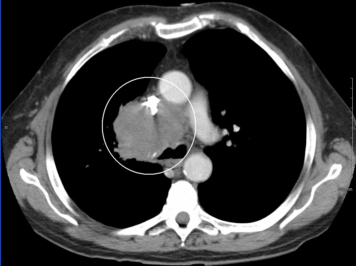 CT
CT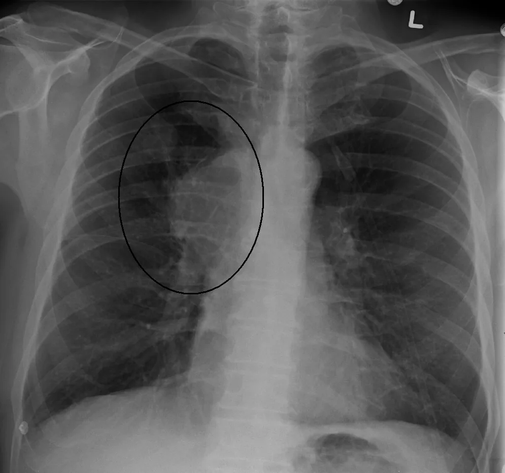 X-ray
X-ray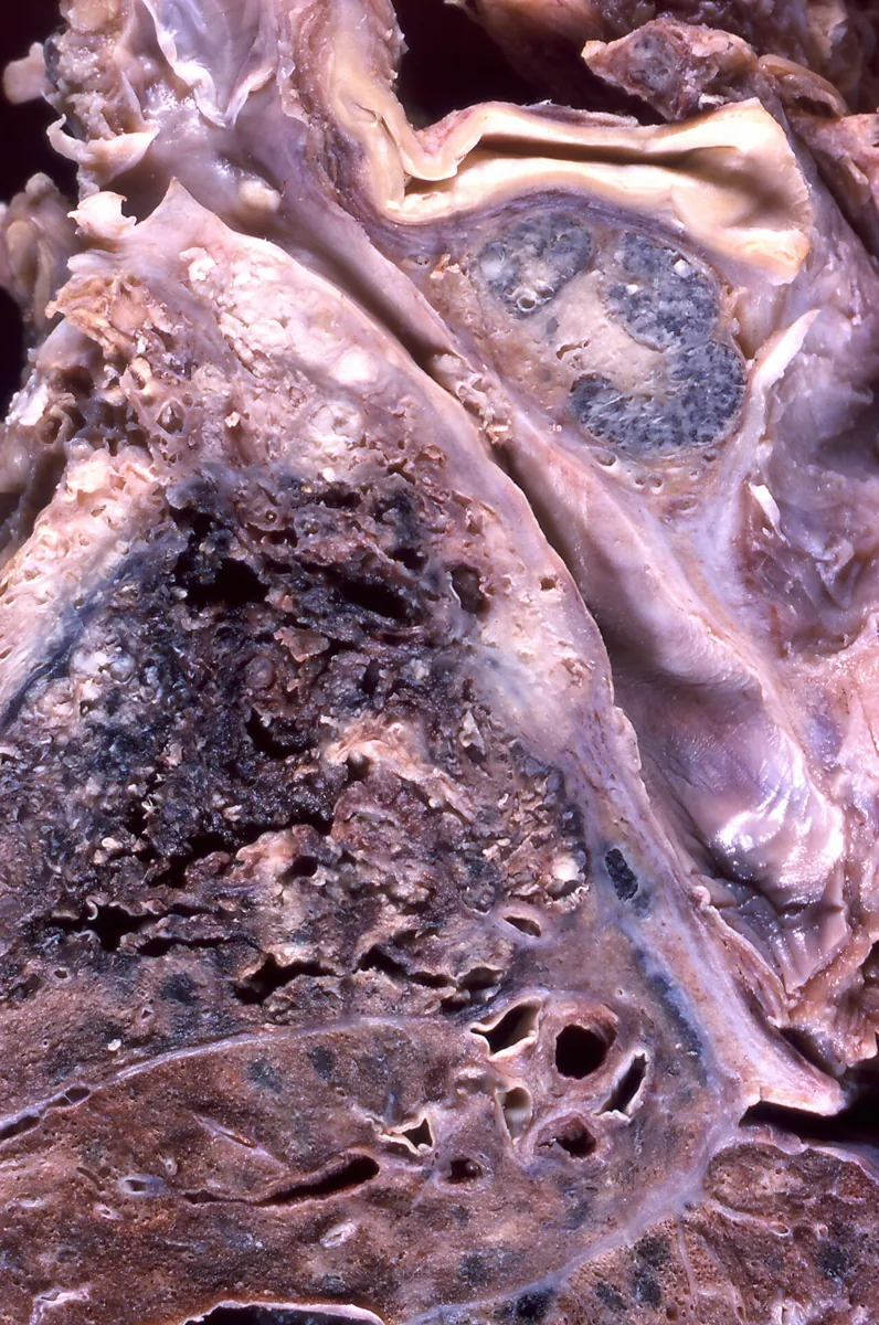 Gross specimen
Definition
Superior vena cava syndrome comprises the symptoms and findings resulting from narrowing or occlusion of the thin-walled superior vena cava. Venous pressure in the head, neck, arms, and upper trunk rises; collateral veins can form an alternative venous pathway.
Causes: About 70 % are tumor-related, due to external compression, ingrowth of tumor cells into the vessel, or thrombosis. Lung carcinoma and non-Hodgkin lymphoma together cause about 85 % of tumor-related cases (non-small cell lung carcinoma about 50 %, non-Hodgkin lymphoma about 10 %); the rest is mainly due to mediastinal metastases, e.g., of breast cancer, and rarely germ cell tumors, thymomas, or mesotheliomas. Superior vena cava syndrome may be the first sign of a previously unknown tumor in up to 60 %. Non-tumor causes (up to about 30 %) are mainly thromboses on central venous catheters and pacemaker leads, as well as mediastinal fibrosis, retrosternal goiter, and Behçet disease.
Clinical features
Symptoms usually develop over weeks to months. Severity and pace depend on the location, speed, and degree of obstruction and on the collaterals.
- Common: swelling of the face and neck, breathlessness (often worse when supine), a sensation of head fullness, cough, distended neck and chest wall veins, arm edema, facial flushing (plethora), dizziness on bending forward
- Less common and signs of severe disease: cyanosis of the upper body, hoarseness, stridor due to laryngeal edema, headache, visual disturbances, confusion, and syncope due to cerebral edema
Severity grades
The grading by Yu and colleagues describes severity:
- Grade 0: radiographic occlusion without symptoms
- Grade 1 (mild): edema of the head or neck, cyanosis, plethora
- Grade 2 (moderate): additional functional impairment, e.g., mild dysphagia, cough, visual disturbances due to eyelid edema
- Grade 3 (severe): mild to moderate cerebral or laryngeal edema or diminished cardiac capacity (syncope after bending forward)
- Grade 4 (life-threatening): significant cerebral edema (confusion, obtundation), laryngeal edema with stridor, or hemodynamic compromise (syncope without precipitating factors, hypotension, renal insufficiency)
- Grade 5: fatal
Diagnosis
- History and examination focusing on the typical signs; confirmation requires imaging.
- Chest x-ray: indirect signs such as mediastinal widening, pleural effusion, lung mass, or right hilar lymphadenopathy.
- Contrast-enhanced CT: the most helpful study; shows the extent of obstruction, collateral veins, and the cause (external compression or thrombosis) as well as mediastinal masses and lymph nodes. Collaterals on CT strongly indicate obstruction (sensitivity 92 %, specificity 96 %).
- Duplex ultrasound of the arm veins: thrombi in the subclavian, axillary, and brachiocephalic veins, especially with catheter thrombosis; the superior vena cava itself cannot be visualized directly by ultrasound.
- Conventional venography: gold standard for depicting obstruction, collaterals, and extent of thrombus; MR venography as an alternative.
- Clarification of the cause: histologic confirmation when a tumor is suspected.
Keep learning in the app
Further reading (open access)
Cross-references
More topics: Haematology & oncology
- Anemia (classification and work-up)
- Hemolytic anemias
- Acute myeloid leukemia (AML)
- Iron deficiency anemia
- Non-Hodgkin lymphomas
- Vitamin B12 deficiency and pernicious anemia
- Multiple myeloma
- Paraneoplastic syndromes
- Autoimmune hemolytic anemia (AIHA)
- Febrile neutropenia
- Immune thrombocytopenia (ITP)
- Renal anemia
Note: Learning content for medical education – not a treatment recommendation and no substitute for diagnosis or treatment decisions in individual cases. Treatment and management are deliberately not covered on this page.