Infectious mononucleosis (glandular fever)
Board exam relevance: in 1 of 105 exam reports · rank 181- Synonyms
- glandular fever, mono, kissing disease, EBV infection, Epstein-Barr virus
- Specialty
- Internal medicine · Infectious diseases
- Images
- Blood smear & cytology 1 · Clinical 1 · CT 1
- Last updated
- 10/2026 · Dr. Pascal Bafteh
Contents
Images (3)
 Blood smear & cytology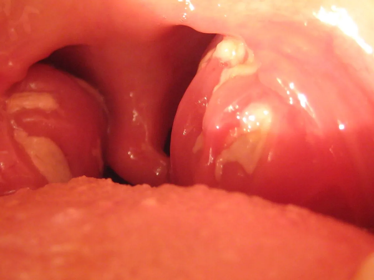
 CT
CTDefinition
Infectious mononucleosis (glandular fever, "kissing disease") is the symptomatic primary infection with Epstein-Barr virus (EBV, human herpesvirus 4). It is characterized by the triad of fever, pharyngitis and lymphadenopathy, often accompanied by marked fatigue that may last for weeks to months.
Occurrence & epidemiology
- EBV infects about half of children before the age of 5; more than 95 % of adults are seropositive, making EBV one of the most widespread human viruses.
- In young children, primary infection is usually asymptomatic; mononucleosis develops mainly in adolescents and adults.
- In lower socioeconomic groups and crowded living conditions, infection more often occurs in early childhood.
Aetiopathogenesis
- Transmission: mainly via saliva through kissing, usually from seropositive people who shed the virus without symptoms; only about 5 % acquire it from someone with acute illness. Rarely via blood products. The virus is not very contagious and is not found in the environment.
- Incubation period: about 30–50 days.
- Pathogenesis: after entry in the oropharynx, EBV infects B lymphocytes. The atypical lymphocytes in blood derive mainly from CD8-positive T cells that eliminate infected B cells by cytotoxicity. Thereafter EBV remains latent for life, mainly in B cells, and is shed intermittently from the pharynx (detectable in 10–20 % of seropositive adults, more often in immunodeficiency).
- Associated diseases: Burkitt lymphoma, certain forms of Hodgkin lymphoma and large B-cell lymphomas, lymphoproliferative disorder after transplantation, nasopharyngeal carcinoma (especially southern China), some cases of hemophagocytic lymphohistiocytosis. X-linked lymphoproliferative syndrome predisposes to severe disease.
Clinical features
Symptoms
- Fever: usually peaks in the afternoon or evening at around 39.5 °C, sometimes up to 40.5 °C.
- Pharyngitis/tonsillitis: often painful and exudative, resembling streptococcal pharyngitis; palatal petechiae, periorbital edema.
- Lymphadenopathy: usually symmetric, especially anterior and posterior cervical nodes; sometimes the only sign.
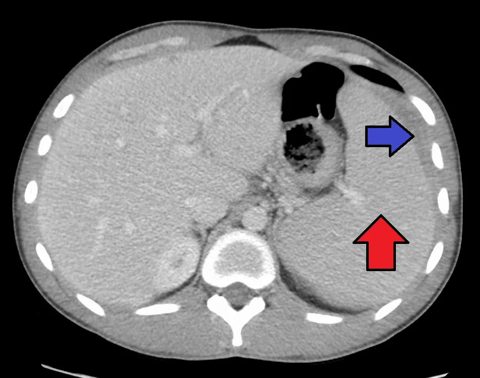
- Splenomegaly in about 50 %, maximal in the 2nd and 3rd week; mild hepatomegaly with percussion tenderness.
- Rash: maculopapular, according to newer data in up to about 20 %.
- Fatigue: most pronounced in the first 2–3 weeks but may last for months.
Complications
- Liver: transaminase elevation to about 2–3 times normal in about 90 %, returning to baseline over 3–4 weeks.
- Blood: transient mild granulocytopenia or thrombocytopenia in about 50 %; hemolytic anemia due to cold agglutinins (anti-i).
- Splenic rupture: most likely 10–21 days after presentation (when splenic enlargement and capsular swelling are maximal), preceded by trauma in only about half of cases; usually painful, rarely presenting only with hypotension.
- Airway: rarely obstruction of the upper airway due to pharyngeal and lymph node swelling.
- Nervous system (rare): encephalitis (including cerebellar), seizures, Guillain-Barré syndrome, peripheral neuropathy, meningitis, myelitis, cranial nerve palsies, psychosis.
- Chronic active EBV infection (rare): fever, interstitial pneumonitis, pancytopenia, hepatitis or uveitis.
Histology
Blood smear
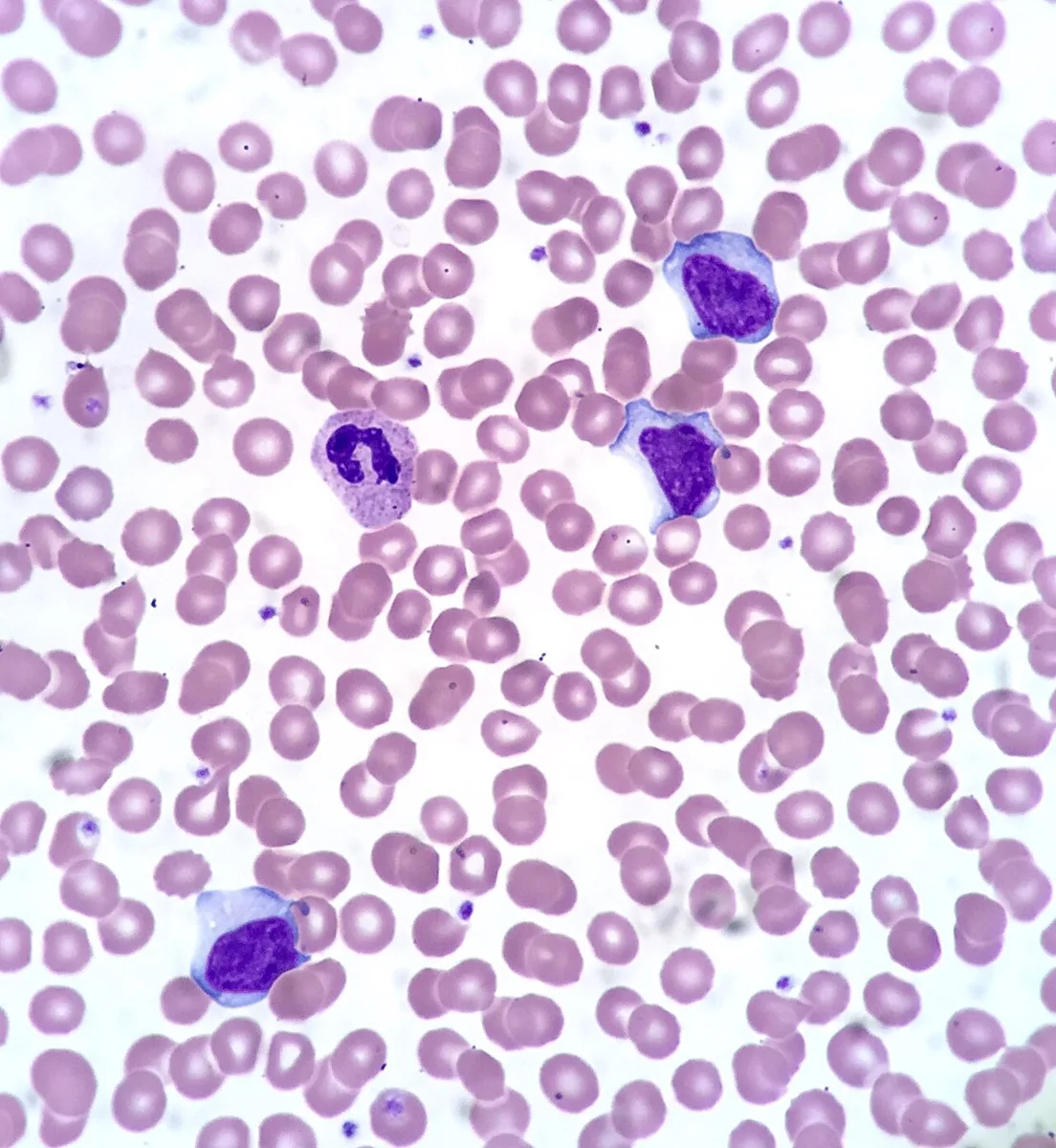
The blood smear shows abundant atypical (reactive) lymphocytes, up to about 30 % of white blood cells. Unlike leukemic cells, they are heterogeneous in appearance. Very high proportions of atypical lymphocytes are seen almost only in primary EBV and CMV infection.
Diagnosis
- Clinical suspicion: posterior cervical or generalized lymphadenopathy and hepatosplenomegaly favor mononucleosis over streptococcal pharyngitis; detection of streptococci in the throat does not exclude mononucleosis.
- Complete blood count: lymphocytosis with atypical lymphocytes.
- Heterophile antibodies (rapid test, "monospot"): positive in 85–90 % of adolescents and adults but in only about 50 % of children aged 2–5 years; titers rise during the 2nd and 3rd week of illness, so an early negative result is repeated after about 7 days. False-positive results in acute HIV infection are possible.
- EBV-specific serology: VCA IgM indicates primary infection (disappears within 3 months); VCA IgG develops early and persists for life; anti-EA IgG appears in the acute phase and falls after 3–6 months; EBNA IgG develops only after 2–4 months and persists for life. Detectable EBNA IgG therefore argues against recent infection.
- Liver tests: transaminases usually moderately elevated; with jaundice or higher elevations, other causes of hepatitis are sought.
- Further tests: with risk factors, HIV RNA and combined antigen/antibody test, as acute HIV infection can look similar; CMV serology if EBV results are negative.
Keep learning in the app
Further reading (open access)
Cross-references
Note: Learning content for medical education – not a treatment recommendation and no substitute for diagnosis or treatment decisions in individual cases. Treatment and management are deliberately not covered on this page.