Sickle cell disease
Board exam relevance: in 2 of 105 exam reports · rank 142- Synonyms
- sickle cell anaemia, sickle cell anemia, drepanocytosis, HbSS disease, sickle cell trait
- Specialty
- Internal medicine · Haematology & oncology
- Images
- Blood smear & cytology 2
- Last updated
- 10/2026 · Dr. Pascal Bafteh
Contents
Images (2)
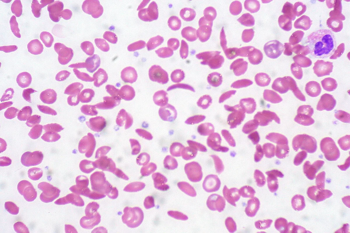 Blood smear & cytology
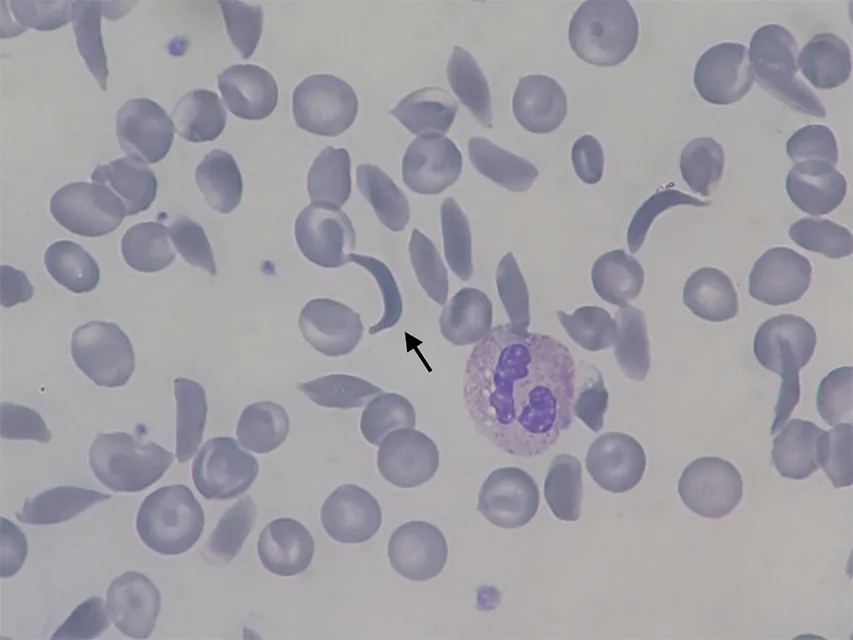 Blood smear & cytology
Definition
Sickle cell disease is an inherited hemoglobinopathy with chronic hemolytic anemia and recurrent vaso-occlusion. It is caused by the abnormal hemoglobin S (HbS). Sickle cell disease comprises homozygous sickle cell anemia (HbSS), HbS-β⁰-thalassaemia and compound forms such as HbSC disease, HbS-β⁺-thalassaemia and other rare variants (e.g. HbSD, HbS-O-Arab).
Heterozygous carriers (sickle cell trait, HbAS) have neither anemia nor pain crises.
Occurrence & epidemiology
The disease is common mainly in people of African origin, and also in the eastern Mediterranean, the Middle East, the Arabian Peninsula, India, the Americas and the Caribbean. Worldwide, an estimated 7.74 million people were living with sickle cell disease in 2021. In the USA about 0.3 % of people of African origin are homozygous and about 8 % are carriers. In Germany an estimated at least 3,000–5,000 children and adults were living with sickle cell disease in 2023.
Aetiopathogenesis
A point mutation in the β-globin gene replaces glutamic acid with valine at position 6 of the β chain. Deoxygenated HbS is poorly soluble, polymerises and deforms red cells into rigid sickle cells at low oxygen partial pressure.
- Vaso-occlusion: the inflexible cells adhere to the endothelium, trigger inflammation and vasoconstriction and plug small arterioles and capillaries; infarction occurs in the microcirculation but also in large vessels (e.g. stroke). Endothelial injury promotes thrombosis.
- Hemolysis: the fragile sickle cells are destroyed by mechanical stress in the circulation.
- Bones: chronic compensatory marrow hyperactivity and ischemia deform the bones.
- Modifying factors: a high HbF fraction (above 10 %) reduces pain crises, acute chest syndrome and leg ulcers; coexisting α-thalassaemia or G6PD deficiency also influence the course.
Crises often occur without an obvious cause, sometimes triggered by fever, viral infections or local trauma.
Clinical features
Chronic findings
- usually compensated anemia of variable severity with pallor and mild jaundice
- in children frequently hepatosplenomegaly; in adults the spleen is usually shrunken by repeated infarction (autosplenectomy) – resulting in increased susceptibility to pneumococcal and Salmonella infections (including Salmonella osteomyelitis)
- cardiomegaly and systolic flow murmur, gallstones, punched-out ulcers around the ankles
- organ damage from repeated ischemia: stroke, moyamoya, seizures, avascular necrosis of bone (e.g. femoral head), renal concentrating defect, papillary necrosis, chronic kidney disease, heart failure, pulmonary hypertension, pulmonary fibrosis, retinopathy
- priapism, mainly in young men
Acute crises
- Vaso-occlusive pain crisis (most common form): severe pain in long bones, hands and feet, back and joints due to ischemia and infarction
- Acute chest syndrome: sudden fever, chest pain and pulmonary infiltrates, rapidly developing hypoxemia with dyspnea; occurs at all ages, most frequent in childhood
- Aplastic crisis: temporary arrest of erythropoiesis in parvovirus B19 infection
- Splenic sequestration: mainly in children whose spleen is not yet fibrotic; acute pooling of cells in the spleen with an Hb fall of at least 2 g/dl up to hemodynamic instability
- Hepatic sequestration with right upper quadrant pain and rapid liver enlargement; intrahepatic cholestasis with marked hyperbilirubinaemia and coagulopathy
- Acute multiorgan failure, often arising from a severe pain crisis or chest syndrome
Histology
Blood smear
Normocytic red cells with sickle cells (sickle or crescent shaped with pointed ends), codocytes, nucleated red cell precursors and polychromasia; microcytosis suggests coexisting thalassaemia. The bone marrow (not needed for diagnosis) shows erythroid hyperplasia.
Diagnosis
- Blood count and signs of hemolysis: anemia, reticulocytes often 10 % or more, bilirubin raised; ESR low
- Hemoglobin analysis (electrophoresis, isoelectric focusing or HPLC): in homozygotes only HbS with a variable HbF fraction; in carriers more HbA than HbS
- Solubility test for HbS (unreliable in the first months of life)
- DNA analysis for prenatal diagnosis and with unclear genotype; newborn screening by hemoglobin analysis
- In acute crises: blood count, reticulocytes (below 1 % in aplastic crisis), chemistry panel; with chest pain or dyspnea chest X-ray and pulse oximetry, with fever cultures
- X-ray (incidental): widened diploic space with radiating trabeculae, thinned cortex of long bones
- Unexplained hematuria also raises the possibility of sickle cell trait.
Keep learning in the app
Further reading (open access)
Cross-references
More topics: Haematology & oncology
- Anemia (classification and work-up)
- Hemolytic anemias
- Acute myeloid leukemia (AML)
- Iron deficiency anemia
- Non-Hodgkin lymphomas
- Vitamin B12 deficiency and pernicious anemia
- Multiple myeloma
- Paraneoplastic syndromes
- Autoimmune hemolytic anemia (AIHA)
- Febrile neutropenia
- Immune thrombocytopenia (ITP)
- Renal anemia
Note: Learning content for medical education – not a treatment recommendation and no substitute for diagnosis or treatment decisions in individual cases. Treatment and management are deliberately not covered on this page.