Psoriatic arthritis
Board exam relevance: in 1 of 105 exam reports · rank 181- Synonyms
- psoriatic arthropathy, arthritis psoriatica, PsA, CASPAR criteria
- Specialty
- Internal medicine · Rheumatology & immunology
- Images
- Clinical 2 · MRI 2
- Last updated
- 10/2026 · Dr. Pascal Bafteh
Contents
Images (4)
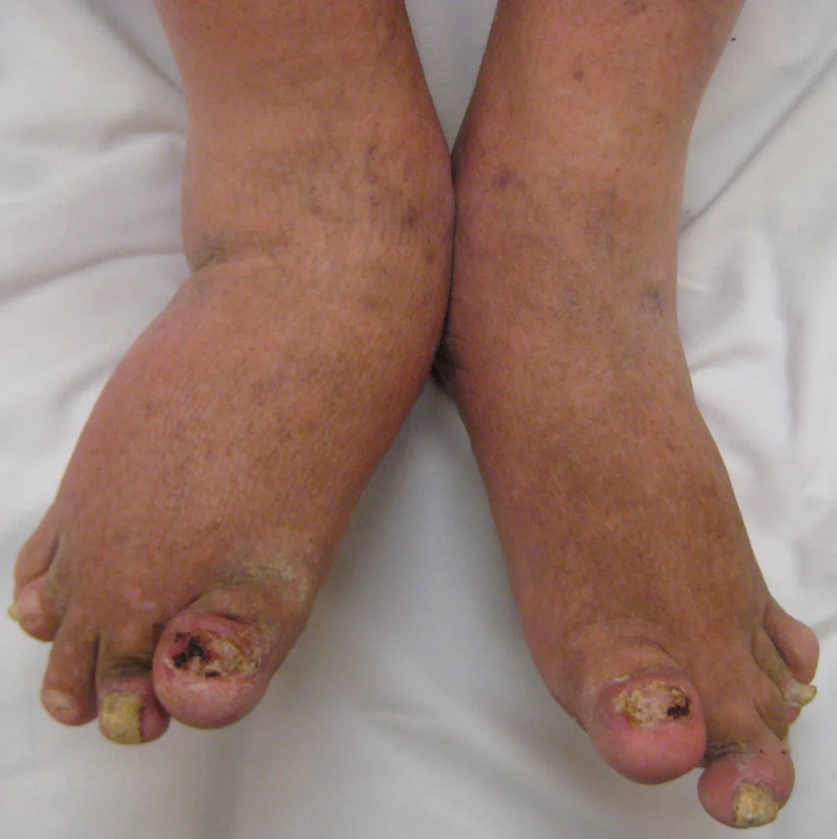
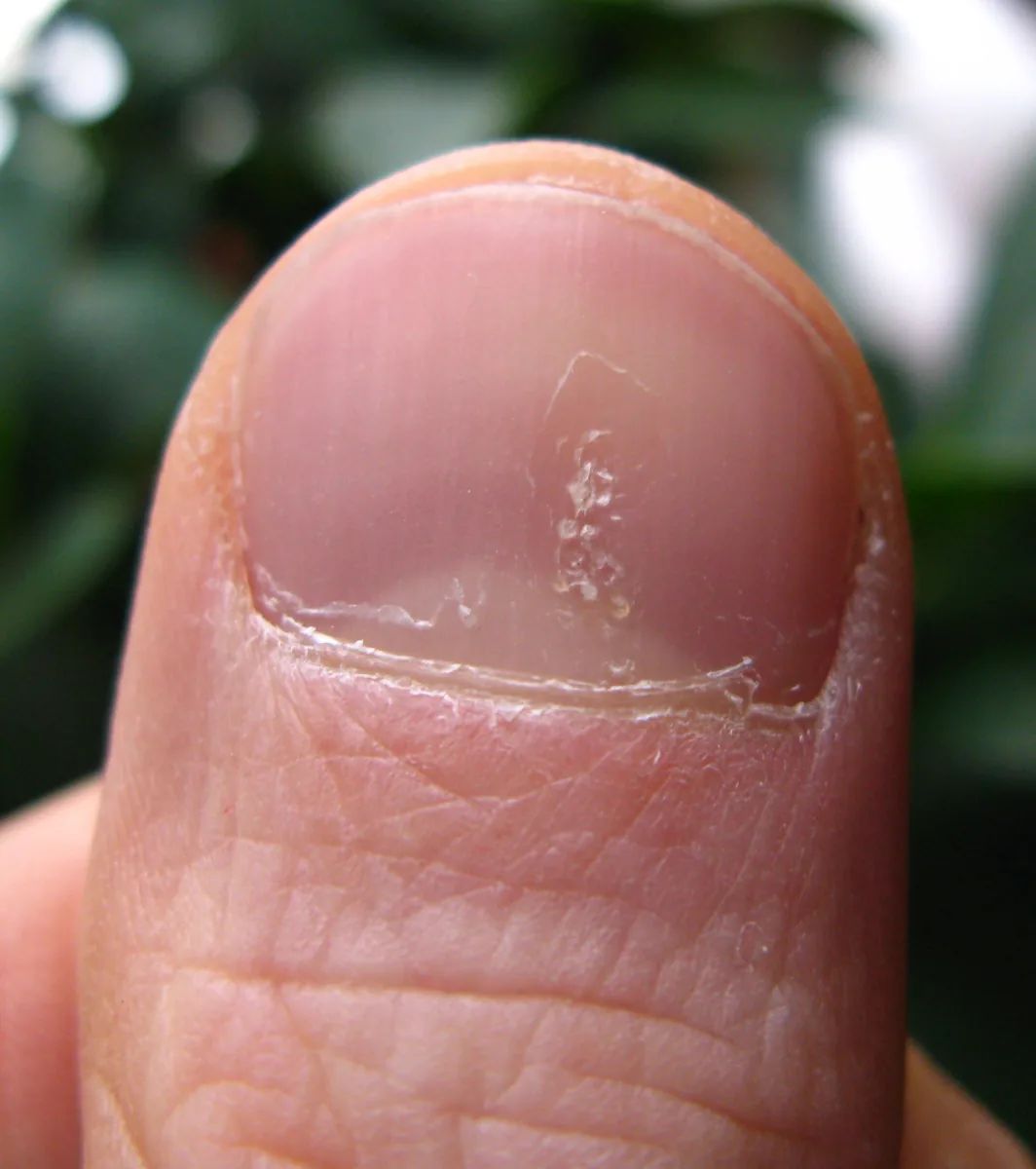
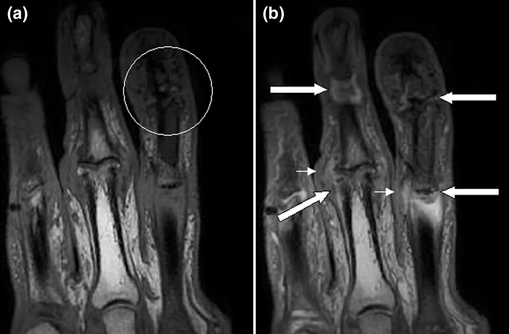 MRI
MRI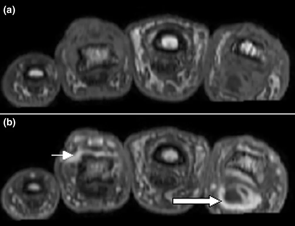 MRI
MRIDefinition
Psoriatic arthritis (PsA) is a chronic inflammatory, seronegative spondyloarthritis in people with psoriasis of the skin or nails. It affects peripheral joints, tendon insertions (entheses), and the axial skeleton. Involvement is often asymmetric and frequently includes the distal interphalangeal joints. The severity of skin and joint involvement is often discordant.
Classification
Subtypes according to Moll and Wright:
- Asymmetric oligoarthritis (up to 4 joints): the most common form at presentation, about 60 %
- Polyarthritis: often symmetric, resembling rheumatoid arthritis
- Distal type with predominant involvement of the distal joints: about 5 %
- Arthritis mutilans: the most severe, rapidly destructive form with bone resorption and telescoping digits
- Axial type with sacroiliitis and spondylitis
CASPAR classification criteria: inflammatory articular disease of joints, spine, or entheses plus at least 3 points from: current psoriasis (2 points) or a personal or family history of psoriasis (1 point); 1 point each for psoriatic nail dystrophy, negative rheumatoid factor, dactylitis (current or documented), juxta-articular new bone formation on radiographs.
Occurrence & epidemiology
PsA develops in about 20 % of people with psoriasis. Prevalence in the general population worldwide ranges from 0.1 to 1 %. PsA is more common in people with HIV infection.
Aetiopathogenesis
Genetic and environmental factors interact. Risk is increased with HLA-B27 and other alleles (HLA-Cw6, HLA-B38, HLA-B39, HLA-DR) in family members. Severe skin and nail psoriasis and uveitis are associated with the development of PsA; physical trauma is also considered a trigger. Skin psoriasis usually precedes the arthritis but may occur simultaneously or only later.
Clinical features
- Involvement of small, medium, and large joints, preferentially the distal interphalangeal joints of fingers and toes.
- Dactylitis (sausage finger or toe) due to inflammation of the flexor tendons; common and asymmetric, in 40–50 %.
- Enthesitis (e.g., Achilles tendon, patellar tendon, epicondyles, spinous processes) in 30–50 %.
- Axial involvement, especially in HLA-B27-positive men, usually as asymmetric sacroiliitis.
- Skin and nails: psoriatic plaques, sometimes hidden on the scalp, ears, gluteal fold, or umbilicus; nail pitting, onycholysis, hyperkeratosis.
- Uveitis as the most common extra-articular manifestation.
- Rheumatoid nodules are absent; skin and joint symptoms may improve or worsen simultaneously.
Diagnosis
- The diagnosis is clinical and requires exclusion of other disorders. In seronegative arthritis with distal joint involvement, asymmetry, lower spine involvement, enthesitis, or dactylitis, psoriasis, nail pitting, and a family history of psoriasis are specifically sought.
- Rheumatoid factor usually negative, occasionally positive; anti-CCP only rarely present.
- Radiographs: distal joint involvement, resorption of terminal phalanges, cupping of proximal phalanges (pencil-in-cup), arthritis mutilans, proliferative new bone formation, enthesitis at the Achilles tendon and plantar fascia insertions.
- Ultrasound detects synovitis, enthesitis, dactylitis, tenosynovitis, and nail changes, often earlier than clinical examination.
Keep learning in the app
Further reading (open access)
Cross-references
More topics: Rheumatology & immunology
- Rheumatoid arthritis
- Systemic lupus erythematosus (SLE)
- Granulomatosis with polyangiitis (GPA)
- Polymyalgia rheumatica
- Gout
- Reactive arthritis
- Behçet's disease
- Giant cell arteritis (temporal arteritis)
- Septic arthritis
- Mixed connective tissue disease (Sharp syndrome)
- Uveitis in rheumatic diseases
- Adult-onset Still's disease
Note: Learning content for medical education – not a treatment recommendation and no substitute for diagnosis or treatment decisions in individual cases. Treatment and management are deliberately not covered on this page.