Behçet's disease
Board exam relevance: in 2 of 105 exam reports · rank 142- Synonyms
- Behcet disease, Behçet syndrome, Adamantiades-Behçet disease, oral aphthae
- Specialty
- Internal medicine · Rheumatology & immunology
- Images
- Clinical 1
- Last updated
- 10/2026 · Dr. Pascal Bafteh
Contents
Images (1)
Definition
Behçet disease is a chronic relapsing multisystem vasculitis with mucosal inflammation. Leading features are recurrent oral ulcers, ocular inflammation, genital ulcers, and skin lesions. In the Chapel Hill nomenclature, it is a variable-vessel vasculitis, as arteries and veins of any size can be affected.
Classification
International criteria: recurrent oral ulcers (at least 3 times in 1 year) plus at least 2 of the following:
- recurrent genital ulcers
- eye lesions (particularly uveitis or retinal vasculitis)
- skin lesions
- positive pathergy test with no other clinical explanation
Occurrence & epidemiology
Men and women are affected about equally, but men often more severely. The disease typically begins in the third decade, occasionally in children. It is most common along the former Silk Road from the Mediterranean to China and uncommon in the United States.
Aetiopathogenesis
The cause is unknown. Immunologic (including autoimmune) mechanisms and viral or bacterial triggers have been suggested. HLA-B51 is a major risk factor; the allele has a prevalence of more than 15 % among people from Europe, the Middle East, and the Far East but is low among people from Africa, Oceania, and South America. Perivascular and endovascular inflammation of arteries and veins leads to thrombosis, aneurysms, pseudoaneurysms, hemorrhage, and stenosis.
Clinical features
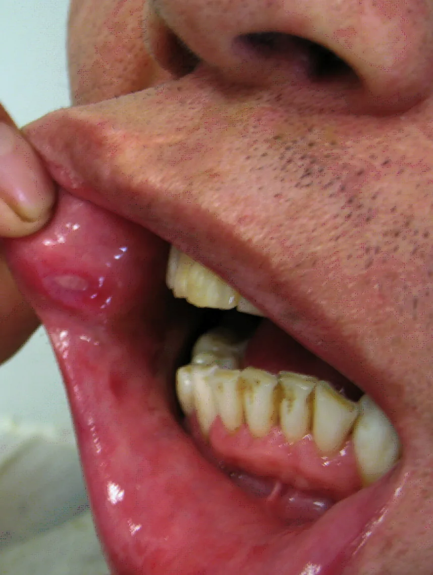
- Oral aphthae in almost all patients, usually the first manifestation: painful, round or oval, 2–10 mm, with a yellowish necrotic center, often in clusters, lasting 1–2 weeks.
- Genital ulcers on the penis and scrotum or vulva (painful) or vagina (often little pain).
- Skin: acneiform lesions, nodules, erythema nodosum, superficial thrombophlebitis, pyoderma gangrenosum-type lesions, palpable purpura; pathergy.
- Eyes in 25–75 %: relapsing uveitis or iridocyclitis with pain, photophobia, and redness, hypopyon; typically bilateral and episodic, often as panuveitis; choroiditis, retinal vasculitis, vascular occlusion, and optic neuritis can lead to blindness.
- Joints in about 50 %: mild, self-limiting, nondestructive arthralgias or arthritis, especially of the knees.
- Vessels: superficial and deep venous thrombosis, including the venae cavae, hepatic veins (Budd-Chiari syndrome), and dural sinuses; arterial aneurysms of the aorta and pulmonary arteries with risk of rupture; clinically recognized large-vessel involvement in 3–5 %.
- Nervous system: parenchymal involvement with pyramidal signs, aseptic meningitis or meningoencephalitis, dural sinus thrombosis; later psychiatric changes.
- Bowel: abdominal pain and diarrhea with ulcers mainly in the ileum and colon, resembling Crohn disease.
Diagnosis
- The diagnosis is clinical and based on the international criteria; it is often delayed because many signs are nonspecific.
- Pathergy test: an erythematous papule larger than 2 mm or pustule 24–48 hours after oblique insertion of a sterile needle into the skin of the forearm is considered positive.
- Laboratory tests: nonspecific signs of inflammation (elevated ESR and CRP, elevated alpha2 and gamma globulins, mild leukocytosis); no specific laboratory test.
- Ophthalmologic examination with slit lamp and fundoscopy; aphthous lesions may show an occlusive vasculitis.
Keep learning in the app
Further reading (open access)
Cross-references
More topics: Rheumatology & immunology
- Rheumatoid arthritis
- Systemic lupus erythematosus (SLE)
- Granulomatosis with polyangiitis (GPA)
- Polymyalgia rheumatica
- Gout
- Reactive arthritis
- Giant cell arteritis (temporal arteritis)
- Septic arthritis
- Mixed connective tissue disease (Sharp syndrome)
- Psoriatic arthritis
- Uveitis in rheumatic diseases
- Adult-onset Still's disease
Note: Learning content for medical education – not a treatment recommendation and no substitute for diagnosis or treatment decisions in individual cases. Treatment and management are deliberately not covered on this page.