Rheumatoid arthritis
Board exam relevance: in 12 of 105 exam reports · rank 20- Synonyms
- rheumatism, chronic polyarthritis, inflammatory arthritis, anti-CCP, rheumatoid factor
- Specialty
- Internal medicine · Rheumatology & immunology
- Images
- Clinical 3 · Histology 1 · X-ray 2 · CT 1
- Last updated
- 10/2026 · Dr. Pascal Bafteh
Contents
Images (7)
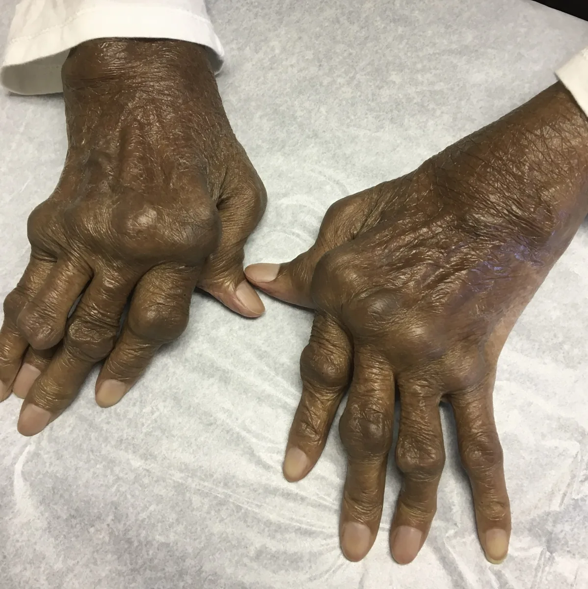
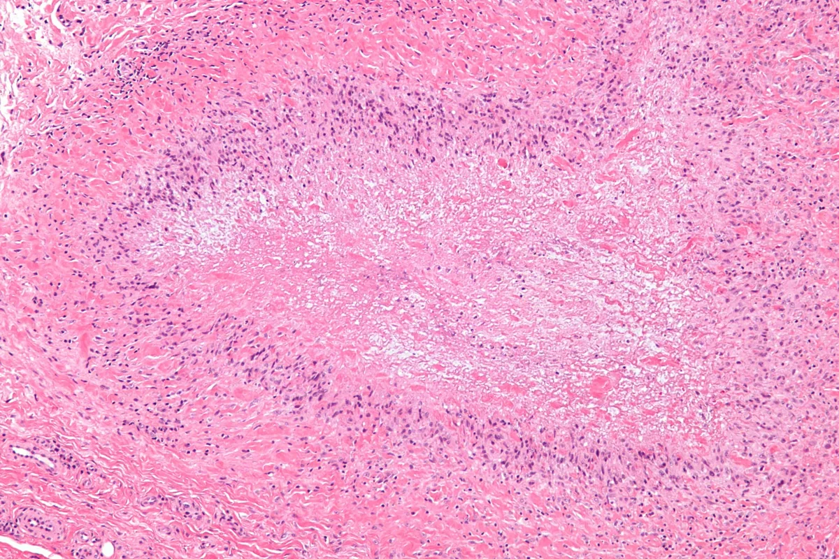 Histology
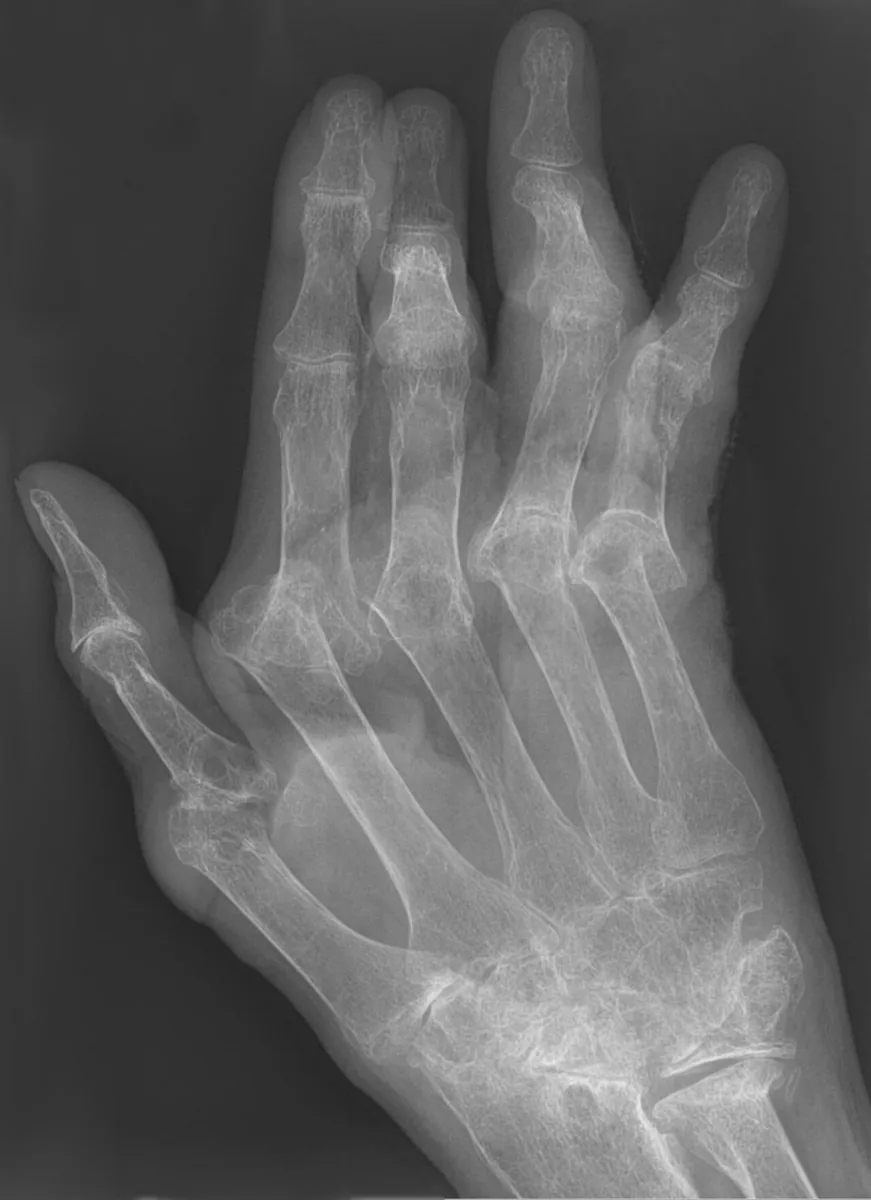 X-ray
X-ray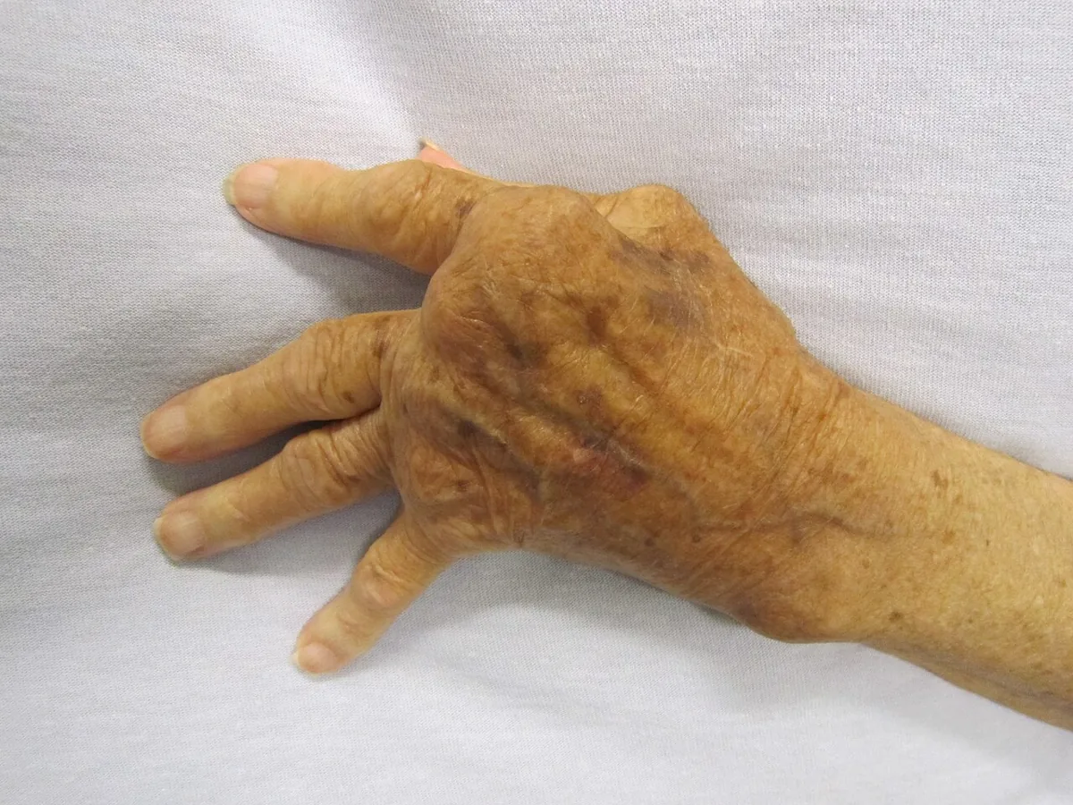
 X-ray
X-ray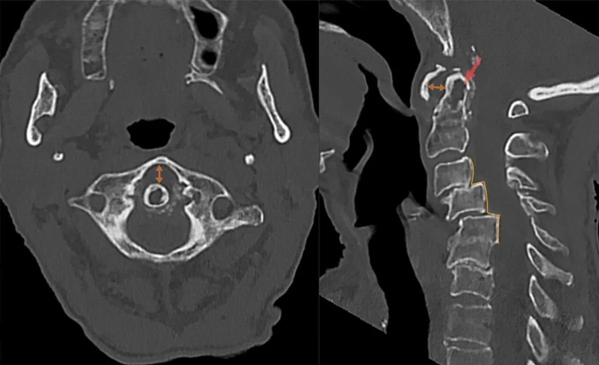 CT
CTDefinition
Rheumatoid arthritis (RA) is a chronic systemic autoimmune disease that primarily involves the joints. It is characterized by symmetric inflammation of peripheral joints, typically the wrists and metacarpophalangeal joints, with progressive destruction of cartilage, bone, and ligaments. Damage is mediated by cytokines and metalloproteases. Systemic symptoms and extra-articular manifestations are common.
Classification
Classification criteria
The 2010 ACR/EULAR classification criteria are used for patients with at least one joint with definite clinical swelling (synovitis) not better explained by another disorder. RA is classified at 6 of 10 points:
- A. Joint involvement: 1 large joint 0; 2–10 large joints 1; 1–3 small joints 2; 4–10 small joints 3; more than 10 joints (at least 1 small joint) 5 points
- B. Serology: rheumatoid factor and anti-CCP negative 0; low-positive (up to 3 times normal) 2; high-positive (more than 3 times) 3 points
- C. Acute-phase reactants: CRP and ESR normal 0; abnormal CRP or ESR 1 point
- D. Duration of symptoms: under 6 weeks 0; at least 6 weeks 1 point
Distal interphalangeal joints, first carpometacarpal joints, and first metatarsophalangeal joints are excluded. Large joints are shoulders, elbows, hips, knees, and ankles; small joints are metacarpophalangeal and proximal interphalangeal joints, metatarsophalangeal joints 2–5, thumb interphalangeal joints, and wrists.
Occurrence & epidemiology
About 0.5 % of the population is affected. Women are affected 2 to 3 times more often than men. Onset may be at any age, most often between 35 and 50 years; the disease also occurs in childhood and old age.
Aetiopathogenesis
The precise cause is unknown. A genetic predisposition is linked to the "shared epitope" in the HLA-DRB1 locus. Risk factors for developing the disease or for greater severity include smoking, obesity, sex hormones, certain drugs, changes in the microbiome of the gut, mouth, and lung, and periodontitis.
Pathogenesis: plasma cells produce autoantibodies such as rheumatoid factor and antibodies against citrullinated peptides (anti-CCP); anti-CCP can often be detected long before the first signs of inflammation. Macrophages and predominantly CD4-positive T cells migrate into the synovium and release tumor necrosis factor-alpha, interleukins, and other mediators. The thickened, villous proliferating synovium (pannus) invades cartilage, subchondral bone, joint capsule, and ligaments; collagenase and stromelysin destroy cartilage, and osteoclastic resorption destroys bone.
Clinical features
Joint involvement
- Usually insidious onset with systemic symptoms (afternoon fatigue, malaise, anorexia, weakness, occasionally low-grade fever) and joint complaints; occasionally abrupt onset that may mimic an acute viral illness.
- Symmetric involvement with pain, swelling, and morning stiffness lasting more than 60 minutes; stiffness also after prolonged inactivity.
- Most commonly involved are the wrists and the metacarpophalangeal joints of the index and middle fingers, as well as proximal interphalangeal joints, metatarsophalangeal joints, shoulders, elbows, hips, knees, and ankles. The distal interphalangeal joints are spared.
- Less common patterns: monoarthritis, polymyalgia-like onset in older adults, palindromic rheumatism.
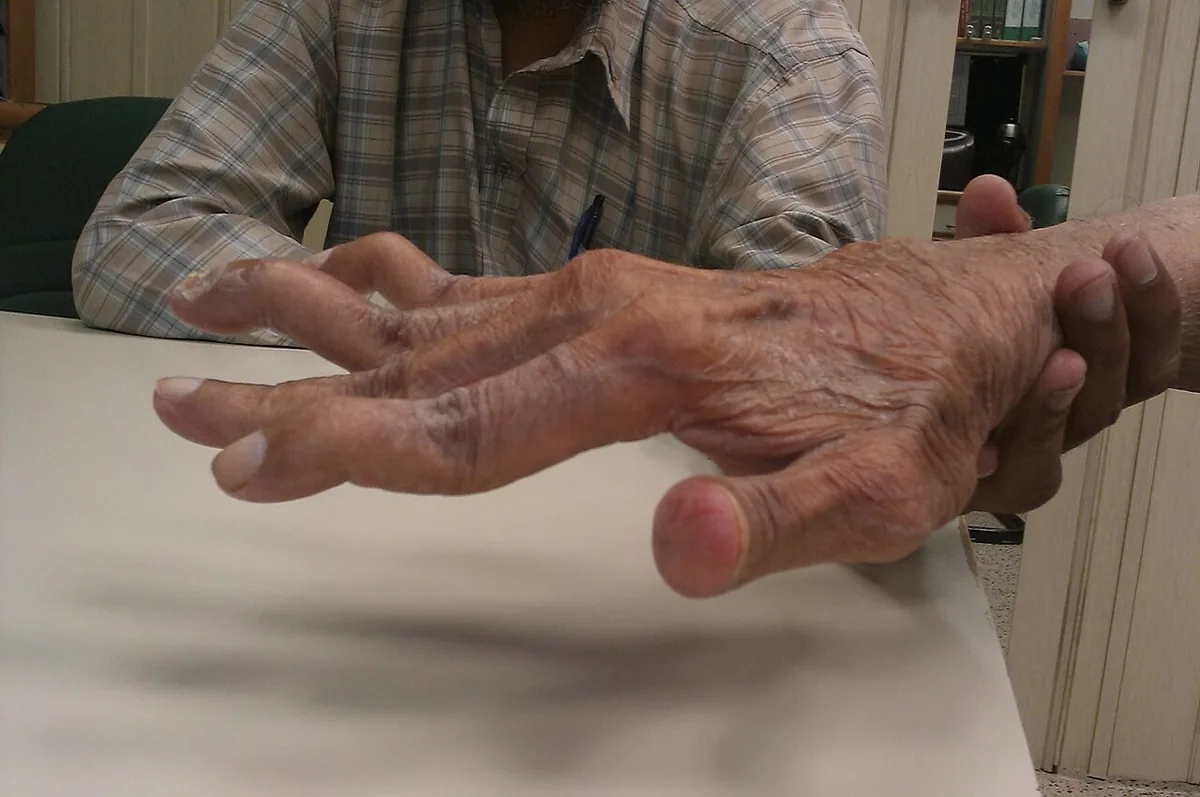
- Deformities: flexion contractures, ulnar deviation of the fingers, swan-neck and boutonnière deformities; carpal tunnel syndrome, Baker cyst.
- Cervical spine: common in long-standing active disease, with possible atlantoaxial subluxation and myelopathy; the lumbar spine is characteristically not involved.
Extra-articular manifestations
- Rheumatoid nodules in up to 30 %, at pressure sites such as the extensor forearm, metacarpophalangeal joints, or soles; also visceral, e.g., in the lung.
- Vasculitis with leg ulcers, digital ischemia, or mononeuritis multiplex.
- Lung and heart: pleural and pericardial effusions, interstitial lung disease, obliterative bronchiolitis, pericarditis, myocarditis.
- Felty syndrome: neutropenia with splenomegaly (in 1–2 %).
- Eyes: secondary Sjögren syndrome, episcleritis, scleromalacia.
- Increased risk of early coronary artery disease, osteoporosis, and certain cancers (lung, lymphoproliferative disorders, non-melanoma skin cancer).
Diagnosis
Laboratory tests
- Rheumatoid factor: in about 70 %; not specific, low titers also in other systemic rheumatic diseases, chronic infections (e.g., hepatitis C), cancers, in 3 % of the general population, and 20 % of older adults.
- Anti-CCP antibodies: specificity about 90 %, sensitivity about 77–86 %; negative in hepatitis C. Rheumatoid factor and anti-CCP do not fluctuate with disease activity.
- ESR and CRP usually elevated in active disease; thrombocytosis.
- Blood count: normochromic, normocytic anemia in up to 60 %; mild polyclonal hypergammaglobulinemia.
- Synovial fluid with acute effusion: turbid, yellow, sterile, usually 10,000–50,000 white cells/µL, no crystals; used to distinguish septic and crystal arthritis.
Imaging and further tests
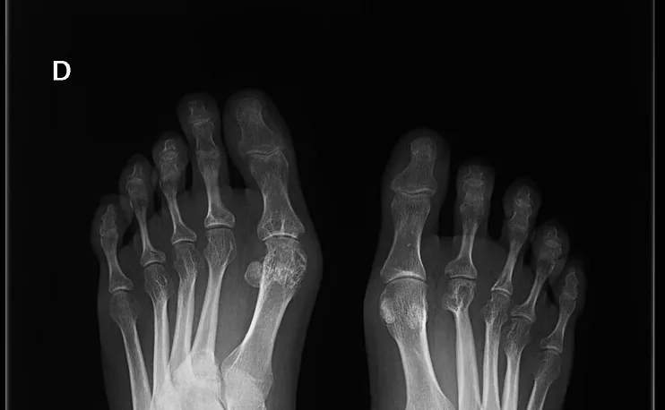
- Radiographs: only soft-tissue swelling in the first months, later periarticular osteoporosis, joint space narrowing, and marginal erosions; erosions often develop within the first year.
- Ultrasound with power Doppler: synovitis, tenosynovitis, and erosions.
- MRI: the most sensitive method for early bone changes.
- Flexion-extension radiographs of the cervical spine when atlantoaxial instability is suspected.
- Pulmonary function tests and high-resolution CT when interstitial lung disease is suspected.
- Activity scores: DAS28 (with ESR or CRP), CDAI, RAPID3.
Keep learning in the app
Further reading (open access)
Cross-references
More topics: Rheumatology & immunology
- Systemic lupus erythematosus (SLE)
- Granulomatosis with polyangiitis (GPA)
- Polymyalgia rheumatica
- Gout
- Reactive arthritis
- Behçet's disease
- Giant cell arteritis (temporal arteritis)
- Septic arthritis
- Mixed connective tissue disease (Sharp syndrome)
- Psoriatic arthritis
- Uveitis in rheumatic diseases
- Adult-onset Still's disease
Note: Learning content for medical education – not a treatment recommendation and no substitute for diagnosis or treatment decisions in individual cases. Treatment and management are deliberately not covered on this page.