Uveitis in rheumatic diseases
Board exam relevance: in 1 of 105 exam reports · rank 181- Synonyms
- iritis, iridocyclitis, eye inflammation, HLA-B27 uveitis, panuveitis
- Specialty
- Internal medicine · Rheumatology & immunology
- Images
- Clinical 1 · Angiography 1
- Last updated
- 10/2026 · Dr. Pascal Bafteh
Contents
Images (2)
 Angiography
Definition
Uveitis is inflammation of the uveal tract, i.e., the iris, ciliary body, and choroid; the retina, anterior chamber, and vitreous are often involved as well. About half of cases are idiopathic. Numerous systemic rheumatic diseases are associated with uveitis, particularly spondyloarthritides, juvenile idiopathic arthritis, sarcoidosis, and Behçet disease.
Classification
By anatomic location:
- Anterior uveitis (iritis, iridocyclitis): e.g., in spondyloarthritides, juvenile idiopathic arthritis, herpesviruses, tubulointerstitial nephritis and uveitis (TINU)
- Intermediate uveitis: e.g., in sarcoidosis, multiple sclerosis, tuberculosis
- Posterior uveitis (retinitis, choroiditis): e.g., in toxoplasmosis, cytomegalovirus, tuberculosis
- Panuveitis: involvement of all segments, e.g., Vogt-Koyanagi-Harada syndrome, Behçet disease
Aetiopathogenesis
- Spondyloarthritides: a common cause of anterior uveitis, most often with ankylosing spondylitis, also with reactive arthritis, inflammatory bowel disease, and psoriatic arthritis; mostly HLA-B27-positive, men more often.
- Juvenile idiopathic arthritis: chronic bilateral iridocyclitis, particularly in the oligoarticular form.
- Rheumatoid arthritis: no isolated uveitis, but scleritis with possible secondary uveitis.
- Sarcoidosis: up to 25 % of all uveitis cases; in 10–50 % of White patients with sarcoidosis, more common in people of African descent and older patients.
- Behçet disease: a common cause of uveitis along the former Silk Road; association with HLA-B51.
- Vogt-Koyanagi-Harada syndrome: suspected autoimmune reaction against melanin-containing cells of the uvea, skin, inner ear, and meninges.
- Rarely, drugs also cause (usually anterior) uveitis.
Clinical features
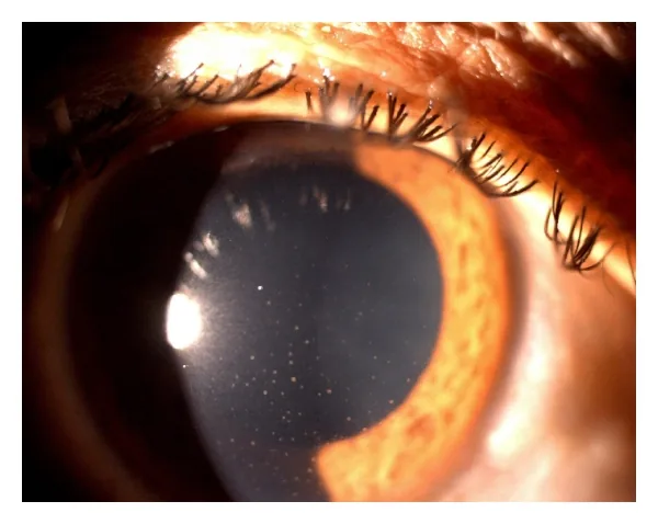
- Anterior uveitis: ocular pain, redness with ciliary flush, decreased vision, and true photophobia; on slit-lamp examination cells and flare in the anterior chamber, keratic precipitates, posterior synechiae, in severe cases hypopyon.
- HLA-B27-associated uveitis: typically unilateral but frequently recurrent, sometimes alternating between eyes.
- Uveitis in juvenile idiopathic arthritis: often without pain, photophobia, and redness ("white iritis"), only with blurred vision and miosis, or entirely asymptomatic.
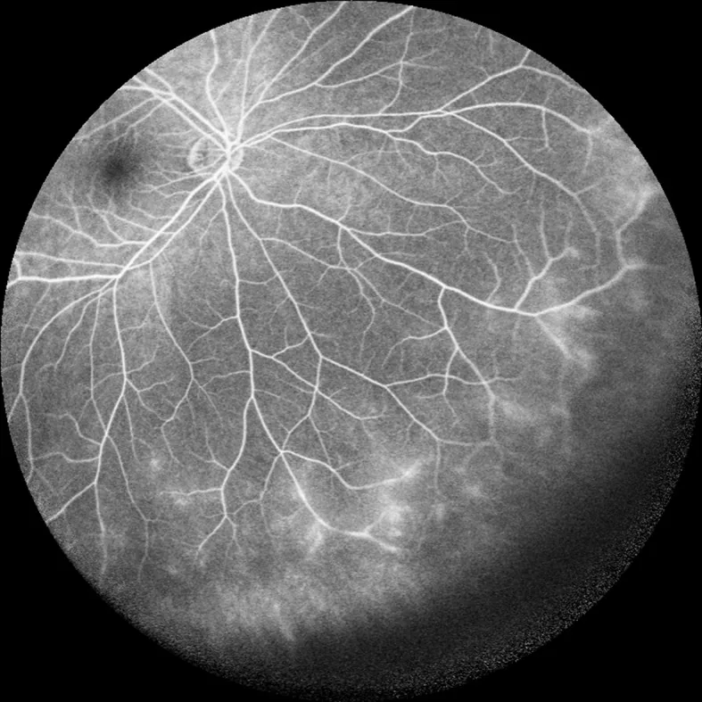
- Intermediate and posterior uveitis: usually painless with floaters and decreased vision; vitreous cells, "snowballs" and "snowbank", cystoid macular edema.
- Sarcoidosis: granulomatous ("mutton-fat") keratic precipitates, granulomas of the conjunctiva, iris, retina, or choroid, retinal vasculitis.
- Behçet disease: severe anterior uveitis with hypopyon, retinitis, retinal vasculitis, optic disk inflammation; frequent recurrences.
- Vogt-Koyanagi-Harada syndrome: early tinnitus, vertigo, headache, and meningismus, later vitiligo, poliosis, and alopecia; serous retinal detachment.
Diagnosis
- Slit-lamp examination: cells and flare in the anterior chamber confirm anterior uveitis.
- Ophthalmoscopy after pupil dilation (indirect more sensitive than direct) for intermediate and posterior findings.
- Intraocular pressure: often (but not always) low in uveitis.
- Search for the cause based on associated findings: e.g., HLA-B27 when spondyloarthritis is suspected, conjunctival biopsy when sarcoidosis is suspected, urine beta-2 microglobulin and possibly renal biopsy in TINU; Behçet disease is diagnosed clinically on the basis of systemic manifestations.
Keep learning in the app
Further reading (open access)
Cross-references
More topics: Rheumatology & immunology
- Rheumatoid arthritis
- Systemic lupus erythematosus (SLE)
- Granulomatosis with polyangiitis (GPA)
- Polymyalgia rheumatica
- Gout
- Reactive arthritis
- Behçet's disease
- Giant cell arteritis (temporal arteritis)
- Septic arthritis
- Mixed connective tissue disease (Sharp syndrome)
- Psoriatic arthritis
- Adult-onset Still's disease
Note: Learning content for medical education – not a treatment recommendation and no substitute for diagnosis or treatment decisions in individual cases. Treatment and management are deliberately not covered on this page.