Septic arthritis
Board exam relevance: in 2 of 105 exam reports · rank 142- Synonyms
- bacterial arthritis, infectious arthritis, joint infection, pyarthrosis, gonococcal arthritis
- Specialty
- Internal medicine · Rheumatology & immunology
- Images
- Clinical 1
- Last updated
- 10/2026 · Dr. Pascal Bafteh
Contents
Images (1)
Definition
Septic (acute infectious) arthritis is a usually bacterial joint infection that evolves over hours or days. The infection resides in the synovium or periarticular tissue and can rapidly destroy joint structures. In adults, gonococcal and nongonococcal forms are distinguished; gonococcal infection is far less destructive to the joint.
Occurrence & epidemiology
Septic arthritis is most common in older adults; half of affected adults are over 60 years of age. In children, about half of those affected are younger than 3 years.
Aetiopathogenesis
Routes of infection: direct inoculation (trauma, bites, arthrocentesis, after surgery on the joint), extension from an adjacent infection (osteomyelitis, soft-tissue abscess), or hematogenous spread.
Pathogens:
- Adults: most often Staphylococcus aureus; also beta-hemolytic streptococci and pneumococci; Neisseria gonorrhoeae in sexually active young adults (slightly over 1 % of cases overall).
- Young children: Kingella kingae is an important pathogen; also streptococci and S. aureus.
- Older and immunocompromised patients: also gram-negative bacteria; Salmonella especially in systemic lupus erythematosus; under immunosuppression also mycobacteria and fungi.
- Bites: often polymicrobial (e.g., Pasteurella multocida after dog and cat bites, Eikenella corrodens after human bites).
Risk factors: older age, rheumatoid arthritis and other damaged joints, previous joint infection, prosthetic joint, injection drug use, diabetes, alcohol use disorder, bacteremia, cancer, immunodeficiency including HIV, immunosuppression, skin infections.
Pathogenesis: neutrophils phagocytose the organizms; their breakdown releases lysosomal enzymes that damage the synovium, ligaments, and cartilage. Articular cartilage can be destroyed within hours or days.
Clinical features
- Nongonococcal bacterial arthritis: within hours to days, moderate to severe joint pain markedly worsened by movement and palpation; effusion, warmth, redness, restricted active and passive motion. In 80 % of adults only one joint is involved, usually the knee, hip, shoulder, wrist, ankle, or elbow; in children, at least 90 % are monoarticular. Fever in most but not all patients.
- Polyarticular involvement more common with immunosuppression, preexisting chronic arthritis, or streptococcal and staphylococcal infection; with injection drug use, often axial joints (sternoclavicular, sacroiliac joints, symphysis pubis).
- Gonococcal arthritis: dermatitis-polyarthritis-tenosynovitis syndrome with fever, multiple skin and mucosal lesions (petechiae, papules, pustules, hemorrhagic vesicles or bullae, necrotic lesions), migratory arthralgias, arthritis, and tenosynovitis, most often in the small joints of the hands, wrists, elbows, knees, and ankles; genital symptoms are often absent.
- Infants and young children: guarding or pseudoparalysis of a limb, irritability, feeding problems, fever or no fever.
- With virulent pathogens, signs of sepsis; under immunosuppression, signs of inflammation are often blunted.
Diagnosis
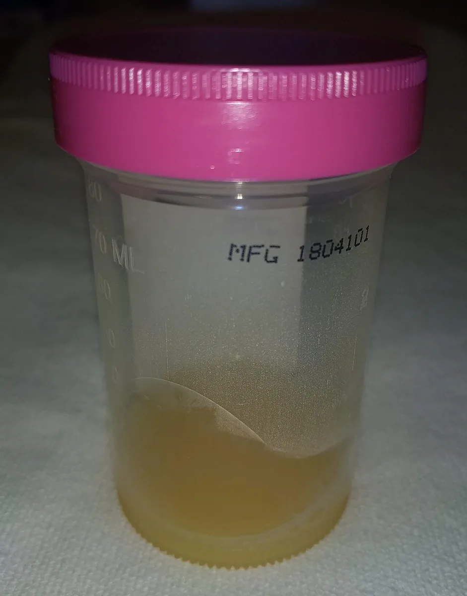
- Arthrocentesis with synovial fluid analysis is the cornerstone: cell count with differential, Gram stain, aerobic and anaerobic culture, crystal analysis. Typical are more than 50,000 white cells/µL (sometimes more than 100,000) with more than 95 % neutrophils; lower counts do not exclude infection early in the course. Foul-smelling fluid suggests anaerobes.
- Gram stain shows organizms in only 50–75 %; culture is definitive. Crystals do not exclude coexisting infection.
- Blood cultures, complete blood count, ESR or CRP; normal results do not exclude infection. Serum urate is not suitable to distinguish gout.
- Molecular tests (PCR, possibly sequencing) on synovial fluid; when gonococcal infection is suspected, nucleic acid amplification tests from cervix, urethra, pharynx, and rectum.
- Imaging: radiographs initially show only soft-tissue swelling and effusion, after 10–14 days joint space narrowing and erosions; gas in the joint suggests E. coli or anaerobes. MRI or ultrasound for joints that are difficult to access and to detect effusions, abscesses, or osteomyelitis.
Keep learning in the app
Further reading (open access)
Cross-references
More topics: Rheumatology & immunology
- Rheumatoid arthritis
- Systemic lupus erythematosus (SLE)
- Granulomatosis with polyangiitis (GPA)
- Polymyalgia rheumatica
- Gout
- Reactive arthritis
- Behçet's disease
- Giant cell arteritis (temporal arteritis)
- Mixed connective tissue disease (Sharp syndrome)
- Psoriatic arthritis
- Uveitis in rheumatic diseases
- Adult-onset Still's disease
Note: Learning content for medical education – not a treatment recommendation and no substitute for diagnosis or treatment decisions in individual cases. Treatment and management are deliberately not covered on this page.