Giant cell arteritis (temporal arteritis)
Board exam relevance: in 2 of 105 exam reports · rank 142- Synonyms
- temporal arteritis, Horton disease, cranial arteritis, GCA, halo sign
- Specialty
- Internal medicine · Rheumatology & immunology
- Images
- Ultrasound 1 · Histology 2
- Last updated
- 10/2026 · Dr. Pascal Bafteh
Contents
Images (3)
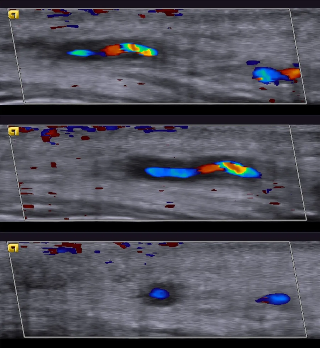 Ultrasound
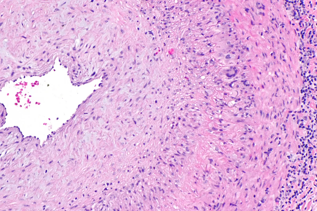 Histology
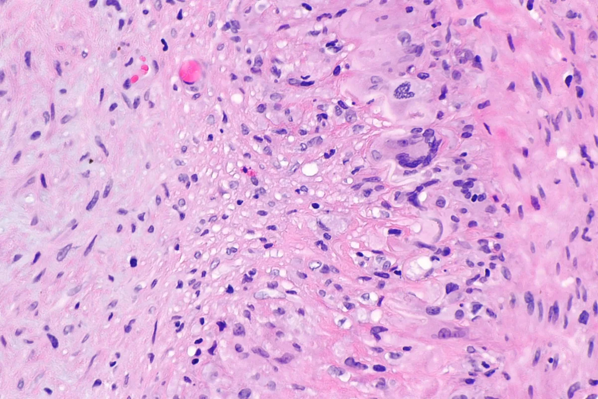 Histology
Definition
Giant cell arteritis (formerly temporal arteritis, Horton disease) is a granulomatous vasculitis of large and medium-sized arteries in older age. It mainly involves the thoracic aorta, the large arteries arising from it in the neck, and extracranial branches of the carotid arteries, especially the temporal artery. In the Chapel Hill nomenclature, it is, like Takayasu arteritis, a large-vessel vasculitis.
Occurrence & epidemiology
Giant cell arteritis is a relatively common form of vasculitis in the United States and Europe; incidence varies by ethnic background. Women are affected more often. The mean age at onset is about 70 years (range 50 to over 90 years). 40–60 % of patients also have polymyalgia rheumatica.
Aetiopathogenesis
The cause is unknown. Inflammation preferentially affects arteries containing elastic tissue and may be localized, multifocal, or widespread; inflamed segments alternate with normal ones (skip lesions). Intimal thickening causes concentric narrowing up to occlusion; involvement of branches of the ophthalmic artery or posterior ciliary arteries causes ischemia of the optic nerve.
Clinical features
- Systemic symptoms: usually low-grade fever, fatigue, malaise, weight loss, sweats; sometimes as fever of unknown origin.
- Headache: the most common symptom, new, severe, sometimes throbbing (temporal, occipital, frontal, or diffuse), with scalp tenderness when combing the hair.
- Claudication of the jaw muscles (especially when chewing firm foods), also of the tongue or extremities.
- Visual disturbances: diplopia, scotomas, ptosis, blurred vision, amaurosis fugax up to sudden, often irreversible vision loss; jaw claudication and diplopia are associated with a higher risk of blindness.
- Neurologic: strokes and transient ischemic attacks.
- Aorta: thoracic aortic aneurysms and dissection as an often late complication.
- Frequently additional symptoms of polymyalgia rheumatica.
Histology
Mononuclear cell infiltrates in the adventitia form granulomas containing activated T cells and macrophages. Multinucleated giant cells, when present, cluster near the disrupted internal elastic lamina. The intima is markedly thickened.
Diagnosis
- Suspicion in people over 50 with new headache, ischemic symptoms above the neck, jaw claudication, temporal artery or scalp tenderness, unexplained fever, or anemia.
- Examination: thickened, tender, sometimes nodular temporal artery with diminished pulse; bruits over neck and arm arteries; fundus with ischemic optic neuropathy (pale, edematous optic disk).
- Laboratory tests: ESR and CRP usually elevated; anemia of chronic disease; sometimes thrombocytosis, mild leukocytosis, low albumin.
- Temporal artery biopsy (segments up to 5 cm increase the yield because of skip lesions); confirms the diagnosis.
- Color Doppler ultrasound of the temporal artery: hypoechoic wall thickening (halo sign) due to wall edema; diagnostic value strongly operator-dependent.
- Imaging of the aorta and its branches to detect large-vessel involvement.
Keep learning in the app
Further reading (open access)
Cross-references
More topics: Rheumatology & immunology
Note: Learning content for medical education – not a treatment recommendation and no substitute for diagnosis or treatment decisions in individual cases. Treatment and management are deliberately not covered on this page.