Primary myelofibrosis
Board exam relevance: in 1 of 105 exam reports · rank 181- Synonyms
- myelofibrosis, idiopathic myelofibrosis, agnogenic myeloid metaplasia, bone marrow fibrosis
- Specialty
- Internal medicine · Haematology & oncology
- Images
- Blood smear & cytology 1 · Histology 1
- Last updated
- 10/2026 · Dr. Pascal Bafteh
Contents
Images (2)
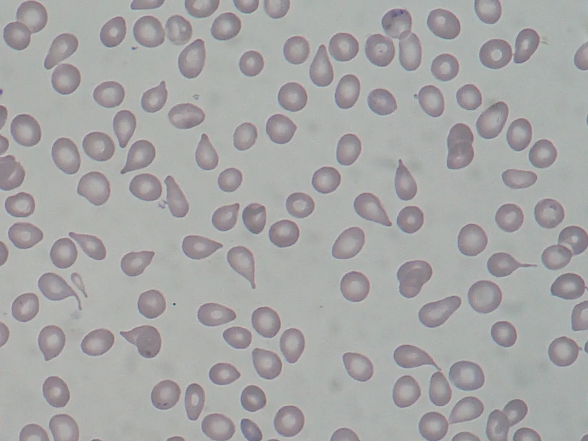 Blood smear & cytology
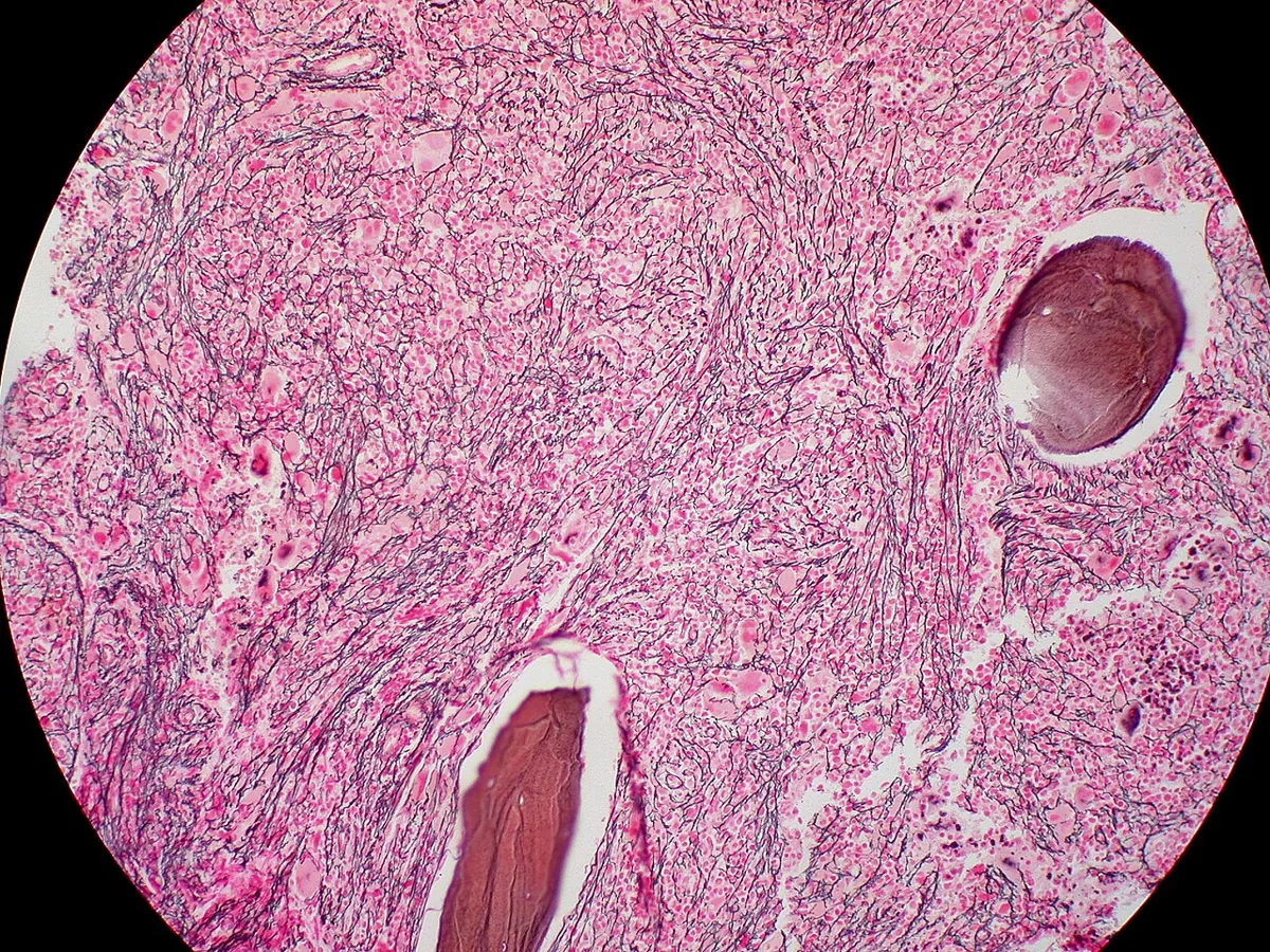 Histology
Definition
Primary myelofibrosis (PMF) is a chronic myeloproliferative neoplasm with bone marrow fibrosis, extramedullary hematopoiesis (mainly in the spleen), splenomegaly, and anemia with nucleated red cell precursors and teardrop-shaped red cells in the blood. The WHO classification distinguishes prefibrotic from fibrotic PMF. Secondary myelofibrosis in other diseases is to be distinguished.
Occurrence & epidemiology
Incidence peaks between 50 and 70 years of age; men are affected more often.
Aetiopathogenesis
The starting point is neoplastic transformation of a pluripotent hematopoietic stem cell with proliferation of atypical megakaryocytes and granulocytes. The progeny of the clone stimulate non-neoplastic fibroblasts to produce excess collagen, resulting in reticulin and collagen fibrosis and osteosclerosis. Driver mutations affect JAK2, CALR, or MPL; rarely none is detectable (triple-negative PMF). Further mutations, particularly in TET2, ASXL1, and DNMT3A, are found in many MPN; less common additional mutations (e.g., in SRSF2, U2AF1, EZH2) are more frequent in PMF than in PV and ET.
Secondary myelofibrosis occurs, among others, with bone marrow metastases, lymphomas, leukemias, multiple myeloma, tuberculosis, benzene or radiation exposure, and following PV or ET.
Clinical features
- Often asymptomatic for a long time.
- Anemia with fatigue and reduced exercise tolerance, increasing over time.
- Splenomegaly, sometimes with splenic infarcts; hepatomegaly in some patients.
- In later stages, malaise, weight loss, and fever.
- Lymphadenopathy is rare.
- Extramedullary hematopoiesis can impair the function of the affected organ.
- Over time, bone marrow failure with thrombocytopenia; transition to acute leukemia is possible.
Diagnosis
- Blood count and smear: anemia; poikilocytosis with teardrop-shaped red cells (dacrocytes); leukoerythroblastic picture with nucleated red cell precursors and immature granulocytes; white cell count usually increased but variable; platelets initially high, normal, or low, later usually decreased.
- LDH frequently elevated.
- Bone marrow biopsy: fibrosis on reticulin or trichrome staining; atypical megakaryocytes.
- Molecular genetics: detection of a JAK2, CALR, or MPL mutation confirms the diagnosis; extended gene sequencing (myeloid panel) detects additional mutations.
- Exclusion of secondary causes of myelofibrosis.
Keep learning in the app
Further reading (open access)
Cross-references
More topics: Haematology & oncology
- Anemia (classification and work-up)
- Hemolytic anemias
- Acute myeloid leukemia (AML)
- Iron deficiency anemia
- Non-Hodgkin lymphomas
- Vitamin B12 deficiency and pernicious anemia
- Multiple myeloma
- Paraneoplastic syndromes
- Autoimmune hemolytic anemia (AIHA)
- Febrile neutropenia
- Immune thrombocytopenia (ITP)
- Renal anemia
Note: Learning content for medical education – not a treatment recommendation and no substitute for diagnosis or treatment decisions in individual cases. Treatment and management are deliberately not covered on this page.