Polycythemia vera
- Synonyms
- polycythemia rubra vera, Vaquez disease, too many red blood cells, myeloproliferative neoplasm
- Specialty
- Internal medicine · Haematology & oncology
- Images
- MRI 1 · Blood smear & cytology 1
- Last updated
- 10/2026 · Dr. Pascal Bafteh
Contents
Images (2)
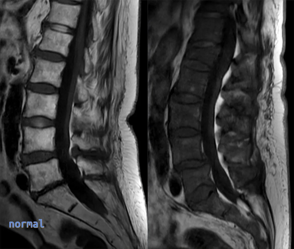 MRI
MRI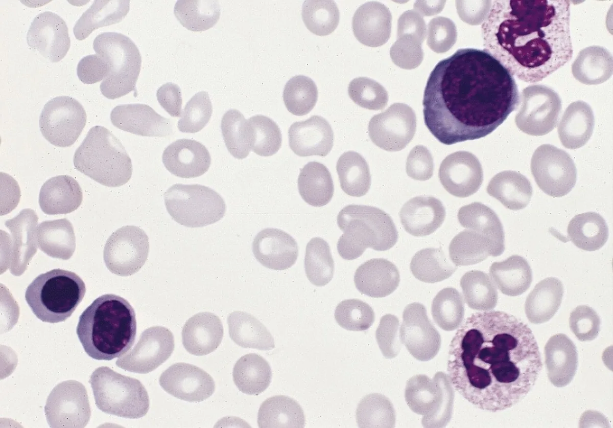 Blood smear & cytology
Definition
Polycythemia vera (PV) is a chronic myeloproliferative neoplasm. Its hallmark is an increase in morphologically normal red blood cells, usually together with increased white cells and platelets (panmyelosis). Erythropoiesis proceeds independently of erythropoietin. Over time, myelofibrosis or, less often, acute myeloid leukemia may develop.
Occurrence & epidemiology
PV is the most common myeloproliferative neoplasm; incidence in the United States is estimated at about 2 per 100,000 and rises with age. The mean age at diagnosis is about 60 years. Women may develop the disease much earlier, sometimes in the second or third decade, often presenting with Budd-Chiari syndrome.
Aetiopathogenesis
The cause is an acquired mutation of a hematopoietic stem cell. The JAK2 V617F mutation (exon 14) is found in about 90 %, JAK2 exon 12 mutations in about 5 %; CALR mutations occur rarely. The mutations permanently activate the JAK2 kinase, which transmits signals from the erythropoietin, thrombopoietin, and G-CSF receptors.
The increased red cell mass raises blood volume and viscosity. An expanded plasma volume may initially mask the erythrocytosis, especially in women. Iron absorption is increased because of reduced hepcidin production; with iron deficiency, a microcytic erythrocytosis develops.
Clinical features
- Often asymptomatic initially; later weakness, headache, light-headedness, visual disturbances, fatigue, and dyspnea.
- Aquagenic pruritus (itching after a hot bath or shower), often the earliest symptom.
- Facial plethora, engorged retinal veins.
- Erythromelalgia: red, warm, painful hands and feet, sometimes with digital ischemia.
- Splenomegaly in more than 30 %.
- Thrombosis, arterial and venous: stroke, deep venous thrombosis, myocardial infarction, retinal vascular occlusion, splenic infarction, Budd-Chiari syndrome; microvascular events such as transient ischemic attacks or ocular migraine.
- Bleeding, mostly gastrointestinal, in about 10 %; with platelets above 1,000,000/µL, acquired von Willebrand syndrome.
- Hyperuricemia with gout and urate stones.
Diagnosis
- Blood count: hemoglobin above 16.5 g/dL in men or above 16.0 g/dL in women raises suspicion. Hematocrit and hemoglobin may be normal because of plasma volume expansion or iron deficiency; the red cell count is the more reliable measure. Neutrophils and platelets are usually increased.
- Microcytic erythrocytosis with a normal hematocrit is typical.
- Molecular genetics: JAK2 V617F and JAK2 exon 12, if negative CALR and LNK.
- Serum erythropoietin: low or low-normal; elevated levels suggest secondary erythrocytosis, which is more common and is excluded first.
- Bone marrow: panmyelosis with large, clustered megakaryocytes, sometimes increased reticulin fibers; not diagnostic.
- Other findings: elevated vitamin B12, hyperuricemia (in at least 30 %); with bleeding, measurement of von Willebrand factor.
- Thrombosis at an unusual site (Budd-Chiari syndrome, portal vein thrombosis) suggests PV.
Keep learning in the app
Further reading (open access)
Cross-references
More topics: Haematology & oncology
- Anemia (classification and work-up)
- Hemolytic anemias
- Acute myeloid leukemia (AML)
- Iron deficiency anemia
- Non-Hodgkin lymphomas
- Vitamin B12 deficiency and pernicious anemia
- Multiple myeloma
- Paraneoplastic syndromes
- Autoimmune hemolytic anemia (AIHA)
- Febrile neutropenia
- Immune thrombocytopenia (ITP)
- Renal anemia
Note: Learning content for medical education – not a treatment recommendation and no substitute for diagnosis or treatment decisions in individual cases. Treatment and management are deliberately not covered on this page.