Polyarteritis nodosa
- Synonyms
- periarteritis nodosa, Kussmaul disease, PAN, DADA2
- Specialty
- Internal medicine · Rheumatology & immunology
- Images
- Histology 1
- Last updated
- 10/2026 · Dr. Pascal Bafteh
Contents
Images (1)
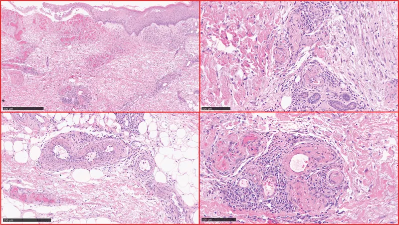 Histology
Definition
Polyarteritis nodosa (PAN) is a systemic necrotizing vasculitis mainly of medium-sized and occasionally small muscular arteries, resulting in secondary tissue ischemia. The kidneys, skin, joints, muscles, peripheral nerves, and gastrointestinal tract are most commonly affected; the lungs are usually spared. In the Chapel Hill nomenclature, it is a medium-vessel vasculitis; ANCA are not typical.
Occurrence & epidemiology
PAN is rare (about 1 case per million). It mainly affects middle-aged adults; incidence increases with age and peaks in people in their 50s.
Aetiopathogenesis
Most cases are idiopathic; immune mechanisms are involved. About 20 % of patients have hepatitis B or C. Drugs may also be a cause. Patients with lymphomas, leukemias, rheumatoid arthritis, or Sjögren syndrome may develop a PAN-like secondary vasculitis. A monogenic, autosomal recessive form is caused by mutations in the adenosine deaminase 2 gene (ADA2 deficiency); VEXAS syndrome (somatic UBA1 mutations) may also present as PAN.
Pathology: segmental, transmural necrotizing inflammation of muscular arteries, preferentially at bifurcations; fresh and older, scarring lesions coexist. Postcapillary venules and veins are not involved, and granulomas are absent. Intimal proliferation with thrombosis leads to infarction, and weakening of the wall to small aneurysms. Glomerulonephritis is not a feature.
Clinical features
- Constitutional symptoms: fever, fatigue, night sweats, loss of appetite, weight loss, weakness; myalgias with focal ischemic myositis, arthralgias, arthritis.
- Peripheral nerves: asymmetric neuropathy, typically mononeuritis multiplex (peroneal, median, ulnar nerves) with footdrop or wristdrop.
- Kidney: hypertension (sometimes rapidly worsening), oliguria, hematuria and proteinuria without casts; renal infarcts with flank pain, perirenal hematomas from ruptured aneurysms.
- Gastrointestinal: abdominal pain, nausea, vomiting, bloody diarrhea, malabsorption, perforation; aneurysms of hepatic or celiac arteries.
- Skin: livedo reticularis, tender nodules, ulcers, infarction and gangrene of fingers or toes.
- Other: orchitis with testicular pain; headache, seizures, stroke; coronary ischemia, heart failure.
Diagnosis
- Biopsy showing necrotizing arteritis, targeted at clinically involved tissue (skin including deep dermis and subcutaneous fat, sural nerve, muscle); skin punch biopsies often miss the lesions. Electromyography and nerve conduction studies help select the biopsy site.
- Arteriography if no biopsy site is available: typical microaneurysms of medium-sized arteries; MR angiography may miss small aneurysms.
- Laboratory tests nonspecific: leukocytosis (up to 20,000–40,000/µL), proteinuria and microscopic hematuria without red cell casts, thrombocytosis, markedly elevated ESR and CRP, anemia, hypoalbuminemia, elevated immunoglobulins, mildly elevated aminotransferases.
- Hepatitis B and C serology in all patients.
- ANCA, rheumatoid factor, anti-CCP, ANA, complement, cryoglobulins, and ENA help exclude other diseases.
Keep learning in the app
Further reading (open access)
Cross-references
More topics: Rheumatology & immunology
- Rheumatoid arthritis
- Systemic lupus erythematosus (SLE)
- Granulomatosis with polyangiitis (GPA)
- Polymyalgia rheumatica
- Gout
- Reactive arthritis
- Behçet's disease
- Giant cell arteritis (temporal arteritis)
- Septic arthritis
- Mixed connective tissue disease (Sharp syndrome)
- Psoriatic arthritis
- Uveitis in rheumatic diseases
Note: Learning content for medical education – not a treatment recommendation and no substitute for diagnosis or treatment decisions in individual cases. Treatment and management are deliberately not covered on this page.