MALT lymphoma
Board exam relevance: in 1 of 105 exam reports · rank 181- Synonyms
- MALToma, extranodal marginal zone lymphoma, gastric lymphoma, marginal zone lymphoma, EMZL
- Specialty
- Internal medicine · Haematology & oncology
- Images
- Histology 2
- Last updated
- 10/2026 · Dr. Pascal Bafteh
Contents
Images (2)
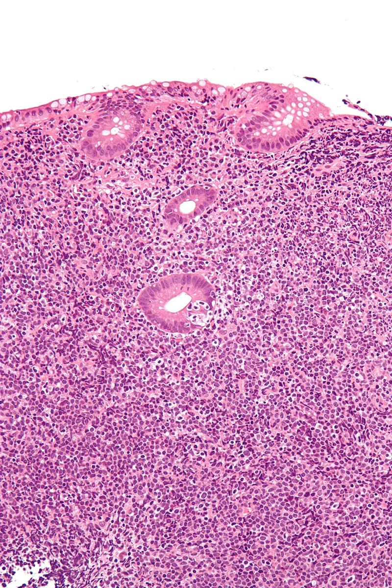 Histology
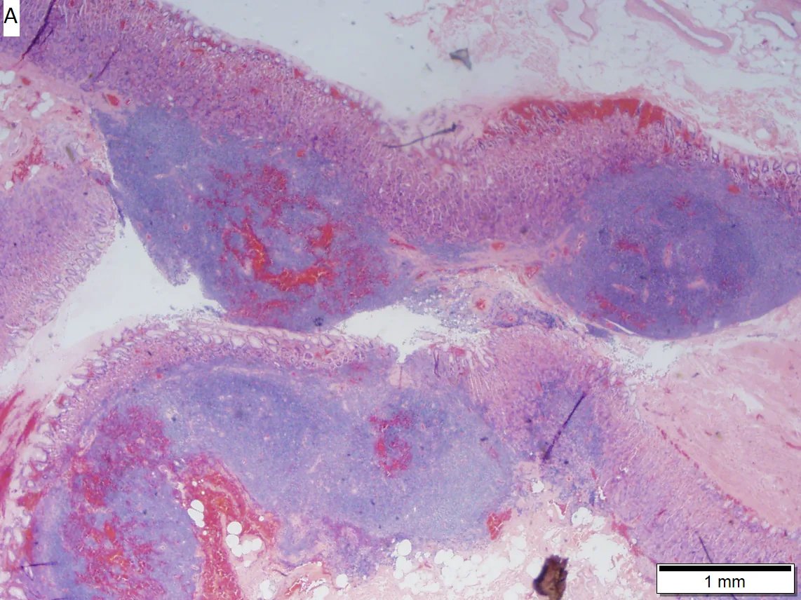 Histology
Definition
MALT lymphoma (extranodal marginal zone lymphoma of mucosa-associated lymphoid tissue) is an indolent B-cell lymphoma. It arises from memory B cells of the marginal zone of secondary lymphoid tissues, usually on the basis of chronic antigenic stimulation by infections or autoimmune disorders. Besides the extranodal type, the WHO classification distinguishes nodal and splenic marginal zone lymphomas.
Occurrence & epidemiology
The most common site is the stomach (about 35 %), followed by the ocular adnexa (13 %), lung (about 9 %), salivary glands (about 8 %), colorectum (5 %), and small intestine (3 %). Gastric MALT lymphoma accounts for 7–9 % of all B-cell lymphomas and 40–50 % of primary gastric lymphomas. It frequently occurs at age 50 to 60. With the decline of Helicobacter pylori infection, the number of new cases is decreasing in some regions.
Aetiopathogenesis
- Helicobacter pylori: the best-documented association; infection leads to accumulation of MALT in the stomach, which normally lacks lymphoid follicles. Tumor-infiltrating T cells recognize H. pylori and activate the B-cell NF-κB pathway via CD40 ligand and BAFF. In the West, the proportion of H. pylori-positive cases has recently declined.
- Other pathogens: Borrelia burgdorferi (skin), Chlamydophila psittaci (ocular adnexa), Campylobacter jejuni (small intestine), hepatitis C virus.
- Autoimmune disorders: Sjögren syndrome, Hashimoto thyroiditis; IgG4-related disease.
- Translocations: t(11;18)(q21;q21) with BIRC3::MALT1 fusion (7–21 % in the stomach, more frequent in H. pylori-negative cases), as well as t(1;14), t(14;18), and t(3;14).
Clinical features
- Stomach: often asymptomatic and an incidental finding at gastroscopy; about 90 % are at stage I at diagnosis.
- Small intestine: abdominal pain, bloating, constipation, or diarrhea; immunoproliferative small intestinal disease (alpha heavy chain disease) causes malabsorption, intermittent diarrhea, and abdominal pain.
- Colorectum: abdominal discomfort, diarrhea, constipation, or a positive fecal occult blood test; preferentially the rectum and ileocecal region.
- Gastric MALT lymphoma carries an increased risk of gastric carcinoma.
Histology
Small to medium-sized, slightly irregular centrocyte-like cells, monocytoid cells, small lymphocytes, and scattered large cells grow in a marginal zone pattern around reactive follicles. Lymphoepithelial lesions (destruction of glandular epithelium by lymphoma cells) are typical, sometimes with plasma cell differentiation and Dutcher bodies. Immunophenotype: CD20+, CD79a+, CD5−, CD10−, CD23−, cyclin D1−; light chain restriction. The Wotherspoon score helps distinguish it histologically from gastritis.
Diagnosis
- Gastroscopy with biopsies: at least 10 biopsies, including from normal-appearing mucosa. Endoscopically, the superficial type predominates (about 70–80 %), which may resemble gastritis or gastric carcinoma; in addition, mass-forming, polypoid, and diffusely infiltrating forms occur.
- Detection of Helicobacter pylori on histology or by noninvasive tests.
- Demonstration of clonality (light chain restriction, PCR for immunoglobulin gene rearrangement) and FISH or RT-PCR for t(11;18)/BIRC3::MALT1.
- Staging: endoscopic ultrasound to assess depth of infiltration and regional lymph nodes; staging according to Lugano (Blackledge) or Paris. FDG-PET detects gastric lesions in only some cases.
Keep learning in the app
Further reading (open access)
- Ishikawa et al., Cancers 2022: MALT Lymphoma in the Gastrointestinal Tract in the Modern Era (PMC-Volltext)
- J Clin Med 2022: Gastric Mucosa-Associated Lymphoid Tissue (MALT) Lymphoma (PMC-Volltext)
- Alaggio et al., Leukemia 2022: WHO-Klassifikation (5. Aufl.), lymphatische Neoplasien (PMC-Volltext)
- MSD Manual Professional: Helicobacter pylori Infection
Cross-references
More topics: Haematology & oncology
- Anemia (classification and work-up)
- Hemolytic anemias
- Acute myeloid leukemia (AML)
- Iron deficiency anemia
- Non-Hodgkin lymphomas
- Vitamin B12 deficiency and pernicious anemia
- Multiple myeloma
- Paraneoplastic syndromes
- Autoimmune hemolytic anemia (AIHA)
- Febrile neutropenia
- Immune thrombocytopenia (ITP)
- Renal anemia
Note: Learning content for medical education – not a treatment recommendation and no substitute for diagnosis or treatment decisions in individual cases. Treatment and management are deliberately not covered on this page.