IgA vasculitis (Henoch-Schönlein purpura)
- Synonyms
- Henoch-Schönlein purpura, HSP, anaphylactoid purpura, IgAV
- Specialty
- Internal medicine · Rheumatology & immunology
- Images
- Clinical 1 · Histology 3
- Last updated
- 10/2026 · Dr. Pascal Bafteh
Contents
Images (4)
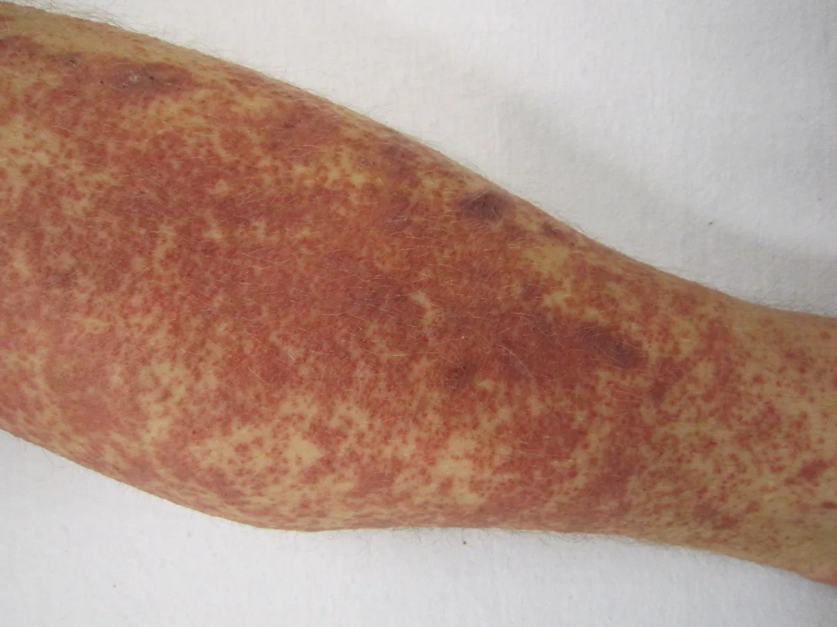
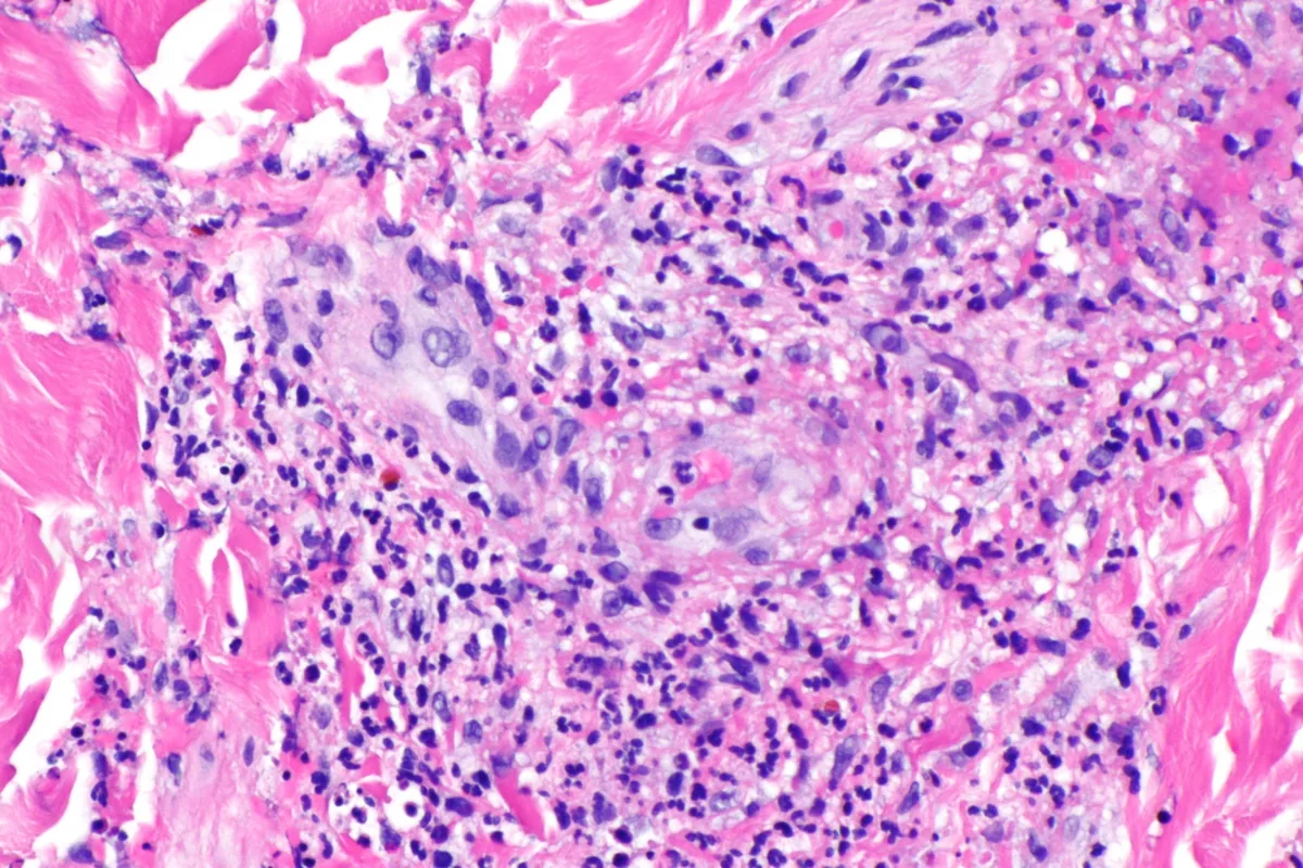 Histology
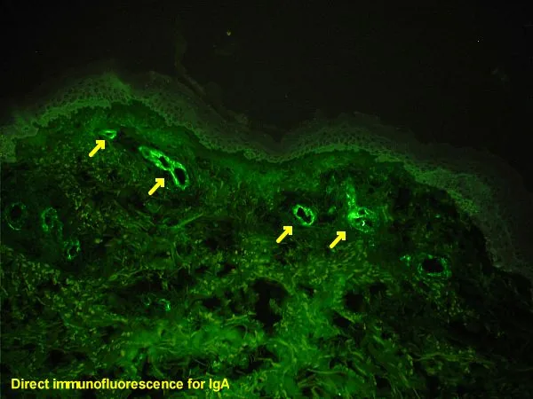 Histology
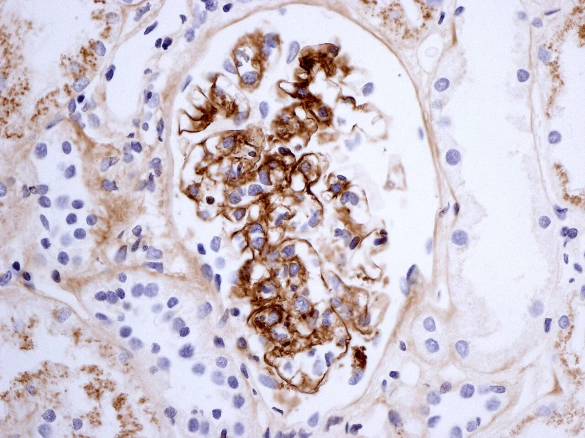 Histology
Definition
IgA vasculitis (formerly Henoch-Schönlein purpura) is a vasculitis affecting primarily small vessels. According to the 2012 Chapel Hill Consensus Conference, it is characterized by IgA1-dominant immune deposits in small vessels of the skin and gastrointestinal tract and frequently causes arthritis. The associated glomerulonephritis is indistinguishable from IgA nephropathy.
Occurrence & epidemiology
IgA vasculitis occurs most often in children. In adults it is less common but more often chronic.
Aetiopathogenesis
IgA-containing immune complexes are deposited in small vessels of the skin and other organs. Possible inciting antigens include viruses causing upper respiratory infections, streptococcal infection, drugs, foods, and insect bites. In the kidney, a focal segmental proliferative, usually mild glomerulonephritis is typical.
Clinical features
- Skin: sudden palpable purpura, typically on the feet, legs, and occasionally the trunk and arms; it may start as urticaria, then becomes palpable, hemorrhagic, and confluent; new crops over days to weeks.
- Joints: fever and polyarthralgias with periarticular tenderness and swelling of the ankles, knees, hips, wrists, and elbows.
- Gastrointestinal tract: colicky abdominal pain, tenderness, melena, positive fecal occult blood test; in children occasionally intussusception.
- Kidney: hematuria, proteinuria, red cell casts.
- Course: symptoms usually subside after about 4 weeks but often recur at least once after a symptom-free interval of several weeks. Rarely, chronic kidney disease develops, more often in adults than in children.
Diagnosis
- In children with a typical picture, the diagnosis is clinical; biopsy is unnecessary when the clinical diagnosis is clear.
- Skin biopsy with direct immunofluorescence (usually part of the work-up in adults): leukocytoclastic vasculitis with IgA deposits in vessel walls.
- Urinalysis: hematuria, proteinuria, and red cell casts indicate renal involvement.
- Complete blood count and renal function tests.
- Renal biopsy when renal function deteriorates; diffuse glomerular involvement or crescents in most glomeruli indicate progressive renal failure.
Keep learning in the app
Further reading (open access)
Cross-references
More topics: Rheumatology & immunology
- Rheumatoid arthritis
- Systemic lupus erythematosus (SLE)
- Granulomatosis with polyangiitis (GPA)
- Polymyalgia rheumatica
- Gout
- Reactive arthritis
- Behçet's disease
- Giant cell arteritis (temporal arteritis)
- Septic arthritis
- Mixed connective tissue disease (Sharp syndrome)
- Psoriatic arthritis
- Uveitis in rheumatic diseases
Note: Learning content for medical education – not a treatment recommendation and no substitute for diagnosis or treatment decisions in individual cases. Treatment and management are deliberately not covered on this page.