Hodgkin lymphoma
Board exam relevance: in 2 of 105 exam reports · rank 142- Synonyms
- Hodgkin disease, Hodgkin's lymphoma, lymph node cancer, classic Hodgkin lymphoma
- Specialty
- Internal medicine · Haematology & oncology
- Images
- Histology 2 · CT 1
- Last updated
- 10/2026 · Dr. Pascal Bafteh
Contents
Images (3)
 Histology
 Histology
 CT
CTDefinition
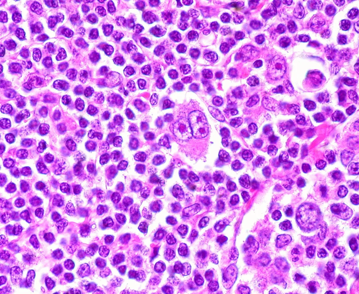
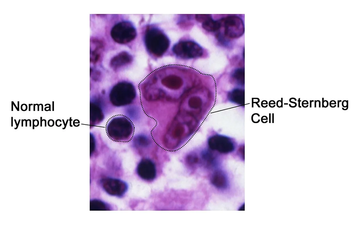
Hodgkin lymphoma (Hodgkin disease, lymphogranulomatosis) is a malignant lymphoma arising from clonally transformed cells of the B-cell lineage. It is characterized by large, binucleated Reed-Sternberg cells in a mixed reactive infiltrate. Lymph nodes, spleen, liver, and bone marrow are mainly involved. Orderly spread to contiguous lymph node regions is typical.
Classification
Classic Hodgkin lymphoma (tumor cells CD15+, CD30+, CD20−) with four histologic subtypes:
- Nodular sclerosis: dense fibrous bands surround nodules of Hodgkin tissue
- Mixed cellularity: a moderate number of Reed-Sternberg cells in a mixed background infiltrate
- Lymphocyte-rich: few Reed-Sternberg cells, many B cells
- Lymphocyte-depleted: numerous Reed-Sternberg cells and extensive fibrosis
Nodular lymphocyte-predominant Hodgkin lymphoma (CD15−, CD30−, CD20+) is now listed as a B-cell lymphoma by the International Consensus Classification.
Stages (Lugano): I one lymph node region or a single extranodal site; II ≥ 2 regions on the same side of the diaphragm; III lymph nodes and/or spleen on both sides of the diaphragm; IV disseminated extranodal involvement (e.g., bone marrow, lung, liver). Suffix A/B for absent or present B symptoms; "bulky" denotes a single mass ≥ 10 cm.
Occurrence & epidemiology
In Germany, about 1,050 women and 1,400 men were diagnosed in 2022. Hodgkin lymphoma is rare but relatively often occurs in young adulthood: between ages 15 and 35, it is among the five most common cancer diagnoses. The age distribution is bimodal, with one peak between 15 and 40 years and a second beyond 60 years; it is rare before age 10.
Aetiopathogenesis
The cause is only partly understood. Discussed factors include:
- Epstein-Barr virus (for some cases) and HIV infection
- congenital immunodeficiencies (e.g., ataxia-telangiectasia, Wiskott-Aldrich syndrome) and immunosuppression, for example in organ transplant recipients
- certain autoimmune disorders (rheumatoid arthritis, celiac disease, Sjögren syndrome, systemic lupus erythematosus)
- familial clustering: children and siblings of affected persons have a slightly increased risk
Many patients develop a slowly progressive defect in cell-mediated immunity.
Clinical features
- Painless lymphadenopathy, mostly cervical; mediastinal lymphadenopathy often an incidental finding on chest x-ray.
- Rarely, pain in involved areas immediately after drinking alcohol.
- B symptoms: fever, night sweats, and unintentional weight loss of more than 10 % of body weight in 6 months; rarely Pel-Ebstein fever (cyclic alternation of febrile and afebrile periods).
- Intense pruritus, often early.
- Splenomegaly common, hepatomegaly unusual.
- Signs of compression: dyspnea and stridor with a mediastinal mass, superior vena cava syndrome, jaundice, lymphedema, Horner syndrome, recurrent laryngeal nerve palsy, spinal cord compression.
- Bone involvement sometimes with osteoblastic vertebral lesions (ivory vertebrae).
Diagnosis
- Lymph node biopsy (preferably excisional biopsy or core needle biopsy; fine-needle aspiration is often inadequate): Reed-Sternberg cells in an infiltrate of histiocytes, lymphocytes, monocytes, plasma cells, and eosinophils; immunohistochemistry (CD15, CD30, CD20).
- Laboratory: complete blood count with differential (slight leukocytosis, lymphocytopenia, eosinophilia, thrombocytosis; anemia in advanced disease), ESR, LDH, alkaline phosphatase, kidney and liver tests.
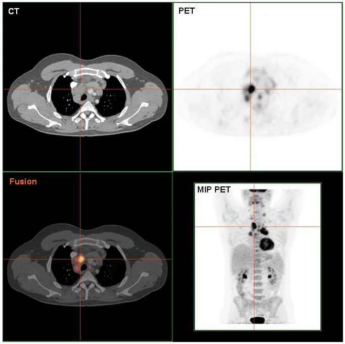
- FDG-PET/CT of chest, abdomen, and pelvis for staging; alternatively contrast-enhanced CT.
- Bone marrow biopsy only if no PET/CT is available.
- MRI in patients with neurologic symptoms.
- Echocardiography and pulmonary function tests as baseline findings.
Keep learning in the app
Further reading (open access)
Cross-references
More topics: Haematology & oncology
- Anemia (classification and work-up)
- Hemolytic anemias
- Acute myeloid leukemia (AML)
- Iron deficiency anemia
- Non-Hodgkin lymphomas
- Vitamin B12 deficiency and pernicious anemia
- Multiple myeloma
- Paraneoplastic syndromes
- Autoimmune hemolytic anemia (AIHA)
- Febrile neutropenia
- Immune thrombocytopenia (ITP)
- Renal anemia
Note: Learning content for medical education – not a treatment recommendation and no substitute for diagnosis or treatment decisions in individual cases. Treatment and management are deliberately not covered on this page.