Hemophilia
Board exam relevance: in 1 of 105 exam reports · rank 181- Synonyms
- bleeder disease, haemophilia A, haemophilia B, factor VIII deficiency, factor IX deficiency, Christmas disease
- Specialty
- Internal medicine · Haematology & oncology
- Images
- X-ray 1
- Last updated
- 10/2026 · Dr. Pascal Bafteh
Contents
Images (1)
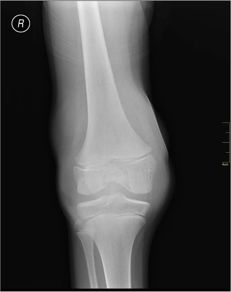 X-ray
X-rayDefinition
Hemophilia is an inherited, X-linked recessive bleeding disorder caused by deficiency of factor VIII (hemophilia A) or factor IX (hemophilia B). Both forms cause the same clinical picture and the same screening test abnormalities and can only be distinguished by specific factor assays. About 80 % of those affected have hemophilia A.
Classification
Severity by residual activity (World Federation of Hemophilia)
- Severe (below 1 %): frequent spontaneous bleeding, especially into joints and muscles; usually diagnosed within the first 2 years of life; genetically mostly large deletions, null mutations or gene rearrangements such as the intron 22 inversion
- Moderate (1–5 %): occasional spontaneous bleeding, prolonged bleeding even after minor injuries; mostly missense mutations
- Mild (5–40 %): bleeding after injuries, after surgery or after dental extractions; without a family history often diagnosed only late in childhood or in adulthood
Normal factor activity is between 50 and 150 %.
Occurrence & epidemiology
Prevalence has traditionally been estimated at about 1:5,000 male live births for hemophilia A and 1:30,000 for hemophilia B; more recent data suggest 1:4,000 and 1:20,000. The German hemophilia registry recorded about 5,400 people with hemophilia A (about 2,900 of them severe) and about 1,000 with hemophilia B in 2022. In about half of newborns with hemophilia there is no family history (new mutation).
Aetiopathogenesis
The cause is mutations, deletions or inversions in the F8 or F9 gene on the X chromosome (more than 3,000 F8 and more than 1,000 F9 variants known). Therefore mainly men are affected. Daughters of a man with hemophilia are obligate carriers; sons of a carrier are affected with a 50 % probability, daughters are carriers with a 50 % probability. Women can also be symptomatic due to skewed X inactivation, usually with factor activity in the range of mild hemophilia.
Pathophysiology: factor VIII acts as the cofactor of activated factor IX in the intrinsic tenase complex, which activates factor X. If either factor is lacking, too little thrombin is generated; the primary platelet plug is not sufficiently stabilised by fibrin. Factor VIII circulates bound to von Willebrand factor, which stabilises it.
Clinical features
The coagulation factor bleeding pattern is typical, with bleeding into deep tissues occurring immediately or delayed after trauma; pain often precedes visible signs of bleeding.
- Hemarthrosis; recurrent joint bleeds lead to synovitis and hemophilic arthropathy
- muscle hematomas, retroperitoneal hemorrhage
- extensive hematomas, rebleeding after surgery and dental extractions
- intracranial hemorrhage even after minor head trauma
- bleeding into the base of the tongue with threatened airway obstruction
- in severe forms onset often in the neonatal period or infancy (e.g. scalp hematoma after delivery), spontaneous bleeding usually in the first years of life
- in women heavy menstrual bleeding and increased bleeding at childbirth
Diagnosis
- History: bleeding pattern and family history; age at first bleeding varies with severity (toddler: spontaneous, child: after injuries, adult: after surgery)
- Global tests: aPTT prolonged (only slightly in mild forms), prothrombin time and platelet count normal; the prolonged aPTT corrects in a mixing study with normal plasma
- Specific factor assays for factor VIII and IX (one-stage aPTT-based or chromogenic assay) – establish type and severity; in non-severe hemophilia A the two assay systems may give discrepant results
- Von Willebrand testing (activity, antigen, multimers) in newly diagnosed, especially mild hemophilia A, since von Willebrand disease (type 2N) also lowers factor VIII
- Molecular genetics: now standard; informs about severity and the risk of neutralising alloantibodies and allows detection of carriers and prenatal diagnosis
- Imaging: ultrasound, X-ray and MRI to assess joint damage
Keep learning in the app
Further reading (open access)
Cross-references
More topics: Haematology & oncology
- Anemia (classification and work-up)
- Hemolytic anemias
- Acute myeloid leukemia (AML)
- Iron deficiency anemia
- Non-Hodgkin lymphomas
- Vitamin B12 deficiency and pernicious anemia
- Multiple myeloma
- Paraneoplastic syndromes
- Autoimmune hemolytic anemia (AIHA)
- Febrile neutropenia
- Immune thrombocytopenia (ITP)
- Renal anemia
Note: Learning content for medical education – not a treatment recommendation and no substitute for diagnosis or treatment decisions in individual cases. Treatment and management are deliberately not covered on this page.