Hemolytic uremic syndrome (HUS)
Board exam relevance: in 3 of 105 exam reports · rank 111- Synonyms
- STEC-HUS, atypical HUS, complement-mediated HUS, hemolytic uremic syndrome
- Specialty
- Internal medicine · Haematology & oncology
- Images
- Histology 1 · Blood smear & cytology 1
- Last updated
- 10/2026 · Dr. Pascal Bafteh
Contents
Images (2)
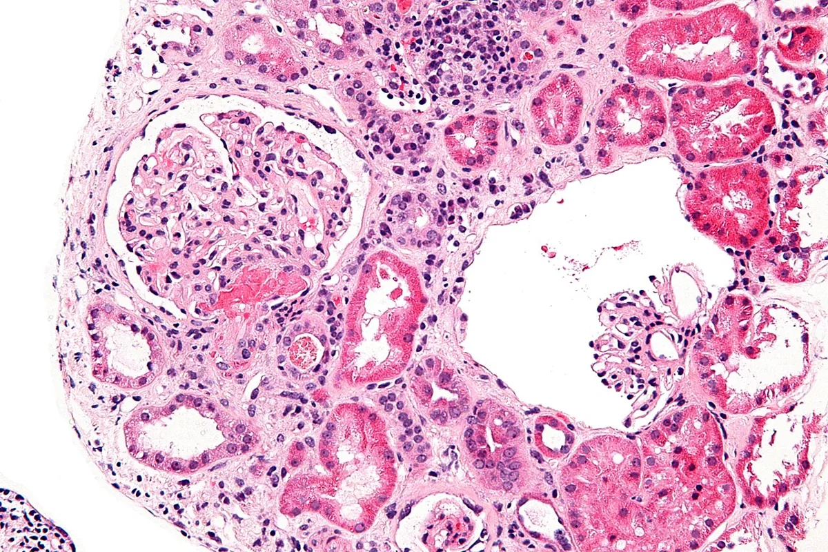 Histology
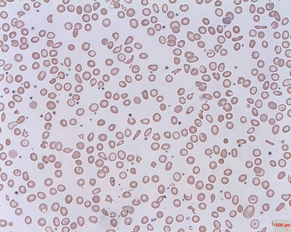 Blood smear & cytology
Definition
Hemolytic uremic syndrome (HUS) is a thrombotic microangiopathy with kidney failure as the leading feature. It is characterised by the triad of Coombs-negative microangiopathic hemolytic anemia, thrombocytopenia and acute kidney injury. Unlike in TTP, ADAMTS13 activity is usually not substantially reduced.
Classification
- STEC-HUS (EHEC-HUS, “typical” HUS): after intestinal infection with Shiga toxin-producing Escherichia coli (e.g. O157:H7) or, less often, Shigella dysenteriae; in children 80–90 % of all HUS cases
- Pneumococcal HUS (SP-HUS): after infection with Streptococcus pneumoniae (pneumonia, meningitis, sepsis); below 5 %
- Complement-mediated HUS (cmHUS, formerly atypical HUS): genetic or acquired dysregulation of the alternative complement pathway; about 5–10 %
- Secondary TMA with an HUS picture, e.g. drug-induced, in autoimmune disease, pregnancy or malignancy
Occurrence & epidemiology
HUS mainly affects children and is the most common cause of acute kidney failure in childhood. It occurs in about 5–10 % of symptomatic EHEC infections in children. Exact figures for adults are lacking. In the largest outbreak to date worldwide, in Germany in 2011 (serotype O104:H4, source contaminated fenugreek sprouts), there were almost 4,000 infections and about 800 HUS cases.
Aetiopathogenesis
All forms share endothelial injury, mainly in the renal capillary bed. In STEC-HUS, Shiga toxin passes from the gut into the bloodstream, binds to the receptor globotriaosylceramide (Gb3) on endothelial cells, is internalised and blocks protein synthesis, leading to cell death. It also activates the alternative complement pathway and is prothrombotic. Severe courses are almost exclusively caused by strains carrying the Shiga toxin gene stx2 (subtype 2a, less often 2c or 2d).
In cmHUS, mutations in regulators such as factor H, factor I or MCP, occasionally autoantibodies against factor H (6–10 %), lead to unrestrained complement activation with endothelial injury. Infections can trigger an episode.
Platelet-rich microthrombi form in the damaged capillaries; they consume platelets and shear red cells (mechanical hemolysis with schistocytes).
Clinical features
STEC-HUS: after an incubation period of usually 2–10 days, a prodrome begins with vomiting, abdominal cramps and frequently bloody diarrhea; fever is usually absent. About 5–13 days after the onset of diarrhea, hemolysis, thrombocytopenia and kidney injury follow.
- Kidney: hematuria, oliguria to anuria, arterial hypertension
- Anemia: pallor, fatigue, jaundice
- Neurological (about one quarter): weakness, confusion, seizures
- Gut: ischemic hemorrhagic colitis with abdominal pain and bloody diarrhea
- Heart: arrhythmias
- purpura or overt bleeding uncommon despite thrombocytopenia
cmHUS: usually without bloody diarrhea, rapidly progressive, often after an upper respiratory tract infection or gastroenteritis; renal involvement ranges from malignant hypertension and proteinuria to acute kidney failure, extrarenal symptoms in about 20 %; tendency to recur.
SP-HUS: in the setting of pneumococcal pneumonia, meningitis or sepsis.
Histology
Blood smear
Schistocytes such as helmet cells and triangular red cell fragments, polychromasia reflecting reticulocytosis and reduced platelets.
Diagnosis
- Blood count and smear: anemia, thrombocytopenia, schistocytes
- Signs of hemolysis: LDH and indirect bilirubin raised, haptoglobin low, reticulocytes raised; direct Coombs test negative
- Renal function and urine: rising creatinine and urea, hematuria, proteinuria
- Coagulation (prothrombin time/INR, aPTT, fibrinogen) largely normal
- ADAMTS13 activity: usually not markedly reduced – the key distinction from TTP
- Pathogen detection with diarrhea: Shiga toxin or stx genes in stool (ELISA, PCR) and culture for EHEC; organizm and toxin may already have cleared at diagnosis, in which case antibodies against O157 lipopolysaccharide in serum can help
- Complement studies (C3, C4, total complement, factor H antibodies, genetic analysis) when cmHUS is suspected; at least half of affected people have no detectable mutation
Keep learning in the app
Further reading (open access)
Cross-references
More topics: Haematology & oncology
- Anemia (classification and work-up)
- Hemolytic anemias
- Acute myeloid leukemia (AML)
- Iron deficiency anemia
- Non-Hodgkin lymphomas
- Vitamin B12 deficiency and pernicious anemia
- Multiple myeloma
- Paraneoplastic syndromes
- Autoimmune hemolytic anemia (AIHA)
- Febrile neutropenia
- Immune thrombocytopenia (ITP)
- Renal anemia
Note: Learning content for medical education – not a treatment recommendation and no substitute for diagnosis or treatment decisions in individual cases. Treatment and management are deliberately not covered on this page.