Essential thrombocythemia
- Synonyms
- primary thrombocythemia, essential thrombocytosis, too many platelets, myeloproliferative neoplasm
- Specialty
- Internal medicine · Haematology & oncology
- Images
- Blood smear & cytology 1 · Clinical 1
- Last updated
- 10/2026 · Dr. Pascal Bafteh
Contents
Images (2)
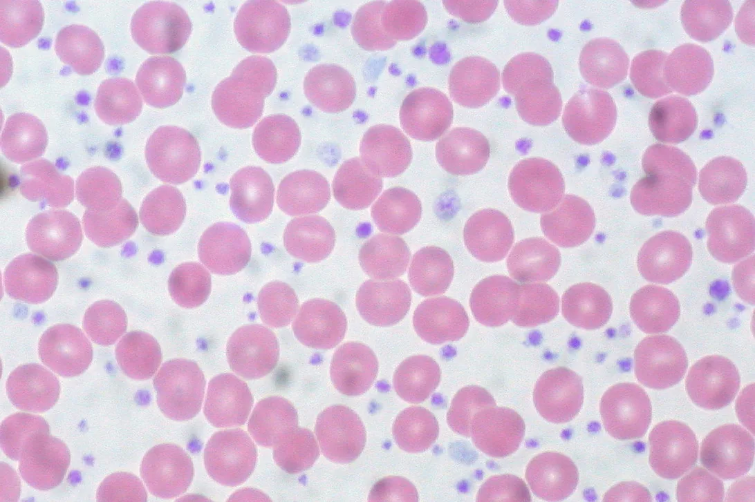 Blood smear & cytology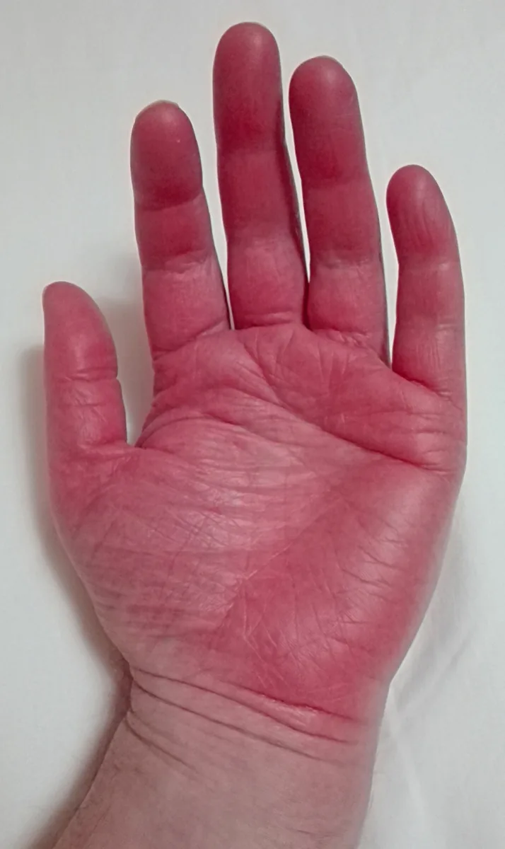
Definition
Essential thrombocythemia (ET) is a myeloproliferative neoplasm with a persistently increased platelet count, megakaryocytic hyperplasia in the bone marrow, and a tendency to bleeding and microvascular circulatory disturbances. It is a diagnosis of exclusion after reactive thrombocytosis and other myeloproliferative neoplasms have been ruled out.
Occurrence & epidemiology
ET usually occurs after age 50; women are affected more often.
Aetiopathogenesis
It is a clonal disorder of the hematopoietic stem cell with increased platelet production. Driver mutations:
- JAK2 V617F in about 50 %
- CALR mutations (exon 9, type 1 and type 2), usually with higher platelet counts
- MPL mutations (thrombopoietin receptor) in a few patients
- Rarely, all three are negative (triple-negative).
With extreme thrombocytosis, platelets adsorb and cleave large von Willebrand multimers, causing type 2 acquired von Willebrand syndrome.
Clinical features
- Microvascular disturbances: erythromelalgia (burning pain, warmth, and redness of the hands and feet, sometimes with digital ischemia and ulcers), headache and ocular migraine, paresthesias, transient ischemic attacks
- Thrombosis, arterial or venous
- Bleeding: usually mild and rarely spontaneous, e.g., epistaxis, bruising, or gastrointestinal bleeding; serious bleeding mainly with extreme thrombocytosis around 1,000,000/µL
- The spleen may be palpable; marked splenomegaly suggests another myeloproliferative neoplasm.
Diagnosis
- Platelet count above 450,000/µL, sometimes above 1,000,000/µL; hematocrit, white cell count, MCV, and iron studies normal.
- Peripheral smear: giant platelets and megakaryocyte fragments.
- Iron studies, because iron deficiency can cause reactive thrombocytosis.
- Molecular genetics: quantitative JAK2 V617F assay and BCR::ABL1 analysis (exclusion of CML); if negative, CALR and MPL. The JAK2 allele burden in ET does not exceed 50 %; higher values suggest PV or PMF.
- Bone marrow biopsy in selected cases: increased numbers of enlarged, mature megakaryocytes.
- Reactive thrombocytosis, CML, masked PV, and myelodysplastic neoplasms with thrombocytosis (e.g., 5q syndrome) are to be distinguished. In about 25 %, a disease initially appearing as ET evolves into overt PV over time.
Keep learning in the app
Further reading (open access)
Cross-references
More topics: Haematology & oncology
- Anemia (classification and work-up)
- Hemolytic anemias
- Acute myeloid leukemia (AML)
- Iron deficiency anemia
- Non-Hodgkin lymphomas
- Vitamin B12 deficiency and pernicious anemia
- Multiple myeloma
- Paraneoplastic syndromes
- Autoimmune hemolytic anemia (AIHA)
- Febrile neutropenia
- Immune thrombocytopenia (ITP)
- Renal anemia
Note: Learning content for medical education – not a treatment recommendation and no substitute for diagnosis or treatment decisions in individual cases. Treatment and management are deliberately not covered on this page.