Disseminated intravascular coagulation (DIC)
Board exam relevance: in 1 of 105 exam reports · rank 181- Synonyms
- consumptive coagulopathy, consumption coagulopathy, defibrination syndrome, disseminated intravascular coagulopathy
- Specialty
- Internal medicine · Haematology & oncology
- Images
- Blood smear & cytology 1
- Last updated
- 10/2026 · Dr. Pascal Bafteh
Contents
Images (1)
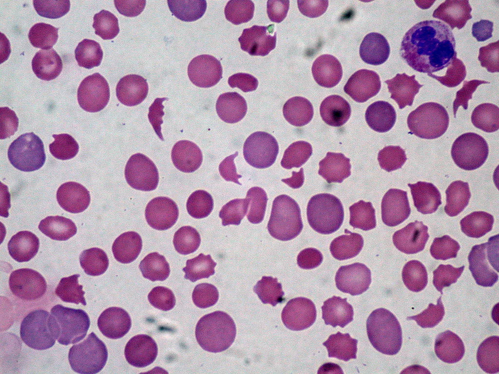 Blood smear & cytology
Definition
Disseminated intravascular coagulation (DIC, consumptive coagulopathy) is an acquired, systemic activation of coagulation with excessive generation of thrombin and fibrin in the circulating blood. Platelets and coagulation factors are consumed in the process. Depending on the course, microthrombosis with organ damage or a bleeding tendency predominates – often both at once. DIC is not a disease in itself but always the consequence of an underlying condition.
Classification
- Acute, rapidly evolving DIC (hours to days): marked consumption of platelets, coagulation factors and fibrinogen with bleeding and microthrombosis up to multiorgan failure; typical in sepsis, trauma, shock and obstetric complications.
- Chronic, slowly evolving DIC (weeks to months): mainly venous thrombosis and embolism, bleeding uncommon; typical in cancer, aortic aneurysms and large hemangiomas.
- Overt and non-overt DIC: the ISTH classification distinguishes manifest, decompensated DIC from still compensated activation of coagulation that becomes apparent only over several measurements.
Aetiopathogenesis
The trigger is usually exposure of the blood to tissue factor. It initiates the extrinsic pathway and thus fibrin formation. Cytokines and disturbed microcirculation cause release of tissue plasminogen activator from the endothelium; the plasmin generated cleaves fibrin into D-dimers and other fibrin degradation products. These in turn impair platelet function. Once platelets and factors are exhausted, bleeding predominates.
Common underlying conditions:
- infections and sepsis, especially due to gram-negative organizms (endotoxin activates tissue factor on monocytes and endothelium)
- obstetric complications: placental abruption, amniotic fluid embolism, retained dead fetus
- cancer, especially adenocarcinomas of the pancreas, stomach, biliary tract, lung, prostate and breast, and acute promyelocytic leukemia
- shock of any cause
Less common: severe tissue damage (head injury, burns, frostbite, gunshot wounds), entry of prostatic tissue after surgery on the prostate, snake venoms, massive intravascular hemolysis (e.g. in ABO incompatibility), aortic aneurysms and cavernous hemangiomas (Kasabach-Merritt syndrome).
Clinical features
- Acute DIC: persistent bleeding from puncture sites and catheter insertion sites, ecchymoses at needle sites, severe gastrointestinal hemorrhage; in addition dysfunction of multiple organs due to microthrombosis and organ bleeding up to multiorgan failure
- Chronic DIC: signs of deep vein thrombosis and pulmonary embolism, occasionally arterial embolism or cardiac valve vegetations; bleeding uncommon
- plus the symptoms of the underlying condition (e.g. sepsis, shock, tumor)
Histology
Blood smear
Thrombocytopenia and, due to mechanical damage on fibrin strands, schistocytes with mild intravascular hemolysis.
Diagnosis
Laboratory tests
- Acute DIC: marked thrombocytopenia, prolonged prothrombin time (low Quick value, raised INR) and aPTT, rapidly falling fibrinogen, markedly raised D-dimer or fibrin degradation products, schistocytes on the smear
- Chronic DIC: mild thrombocytopenia, prothrombin time and aPTT normal or only slightly prolonged (the aPTT is sometimes even shortened initially), fibrinogen normal or moderately reduced, D-dimer raised
- Serial testing: because fibrinogen is raised as an acute-phase protein in many underlying conditions, a fall over two consecutive measurements is more informative than a single value.
- Distinction from severe liver disease: factor VIII is not produced in hepatocytes; it is normal or raised in liver disease but reduced by consumption in DIC. D-dimer levels are usually higher in DIC.
ISTH score for overt DIC
Applicable only in the presence of an underlying condition that can be associated with DIC:
- Platelets: 50 to less than 100 G/l = 1 point; below 50 G/l = 2 points
- Fibrin-related marker (e.g. D-dimer): moderately raised = 2 points; strongly raised = 3 points
- Prolongation of prothrombin time: 3 to less than 6 seconds = 1 point; 6 seconds or more = 2 points
- Fibrinogen: below 1 g/l = 1 point
A total of 5 points or more indicates overt DIC; with lower values, only the trend of repeated measurements detects non-overt DIC.
Keep learning in the app
Further reading (open access)
Cross-references
More topics: Haematology & oncology
- Anemia (classification and work-up)
- Hemolytic anemias
- Acute myeloid leukemia (AML)
- Iron deficiency anemia
- Non-Hodgkin lymphomas
- Vitamin B12 deficiency and pernicious anemia
- Multiple myeloma
- Paraneoplastic syndromes
- Autoimmune hemolytic anemia (AIHA)
- Febrile neutropenia
- Immune thrombocytopenia (ITP)
- Renal anemia
Note: Learning content for medical education – not a treatment recommendation and no substitute for diagnosis or treatment decisions in individual cases. Treatment and management are deliberately not covered on this page.