Chronic myeloid leukemia (CML)
Board exam relevance: in 1 of 105 exam reports · rank 181- Synonyms
- chronic myelogenous leukemia, chronic granulocytic leukemia, Philadelphia chromosome, BCR-ABL, blood cancer
- Specialty
- Internal medicine · Haematology & oncology
- Images
- Blood smear & cytology 3
- Last updated
- 10/2026 · Dr. Pascal Bafteh
Contents
Images (3)
 Blood smear & cytology
 Blood smear & cytology
 Blood smear & cytology
Definition
Chronic myeloid leukemia (CML) is a myeloproliferative neoplasm of the pluripotent stem cell with marked overproduction of mature and immature granulocytes. It is defined by the BCR::ABL1 fusion resulting from the translocation t(9;22)(q34;q11) (Philadelphia chromosome).
Classification
The natural course is biphasic or triphasic:
- Chronic phase: initially indolent; about 85 % are diagnosed in this phase.
- Accelerated phase (former definition): worsening anemia, thrombocytopenia or thrombocytosis, enlarging spleen, basophilia, clonal evolution, and blasts up to 19 %. The 2022 WHO classification no longer includes this phase and instead emphasizes high-risk features of the chronic phase; the 2022 ICC retains it (including 10–19 % blasts or ≥ 20 % basophils in the blood).
- Blast phase: ≥ 20 % myeloid blasts in blood or bone marrow, extramedullary blast proliferation, or increased lymphoblasts. Blasts are myeloid (about 60 %), lymphoid (about 30 %), or megakaryoblastic (about 10 %).
Occurrence & epidemiology
The average age at diagnosis is 66 years. The lifetime risk in the United States is about 0.2 %. CML accounts for about 14 % of newly diagnosed leukemias there.
Aetiopathogenesis
In the translocation t(9;22), the ABL1 gene from chromosome 9 is fused to the BCR gene on chromosome 22. The BCR::ABL1 fusion protein is a constitutively active tyrosine kinase. It increases cell proliferation, reduces adherence of leukemia cells to the bone marrow stroma, and protects them from apoptosis. The clone includes all myeloid lineages plus some T and B cells; hematopoiesis also takes place extramedullarily in the spleen and liver. Ionizing radiation is a known risk factor for leukemias.
Clinical features
- Often asymptomatic initially; frequently an incidental finding on a blood count.
- Nonspecific symptoms: fatigue, weakness, anorexia, weight loss, night sweats, fullness in the left upper quadrant.
- Splenomegaly in 60–70 %, sometimes marked.
- Gouty arthritis due to increased cell turnover.
- Leukostasis at very high cell counts: tinnitus, stupor.
- Pallor, bleeding, fever, marked lymphadenopathy, and skin infiltrates appear only as the disease progresses.
Diagnosis
- Blood count: elevated granulocyte count, usually below 50,000/µL in asymptomatic patients and 200,000 to 1,000,000/µL in symptomatic patients; left shift, basophilia, and eosinophilia; platelets normal or moderately increased; hemoglobin usually above 10 g/dL.
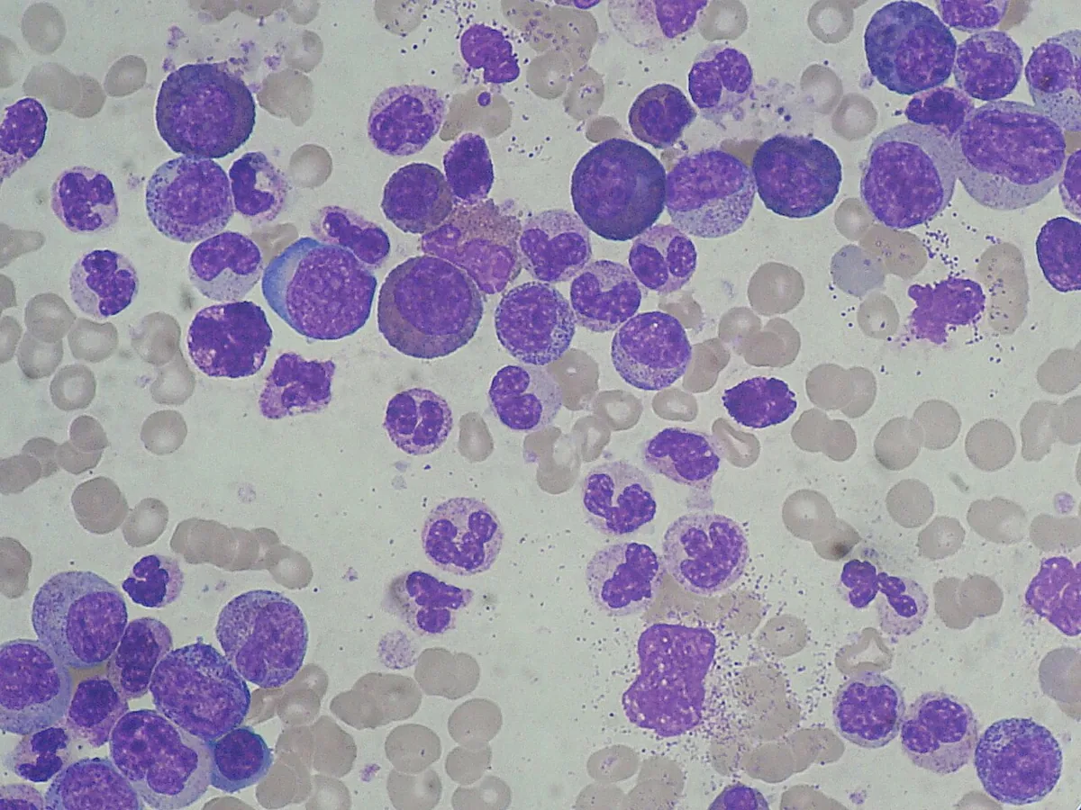
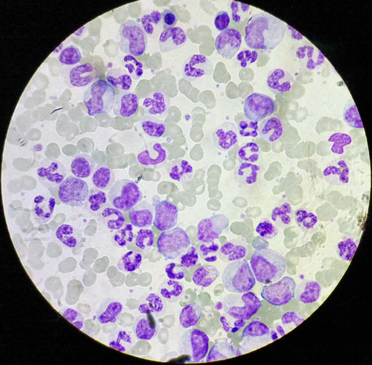
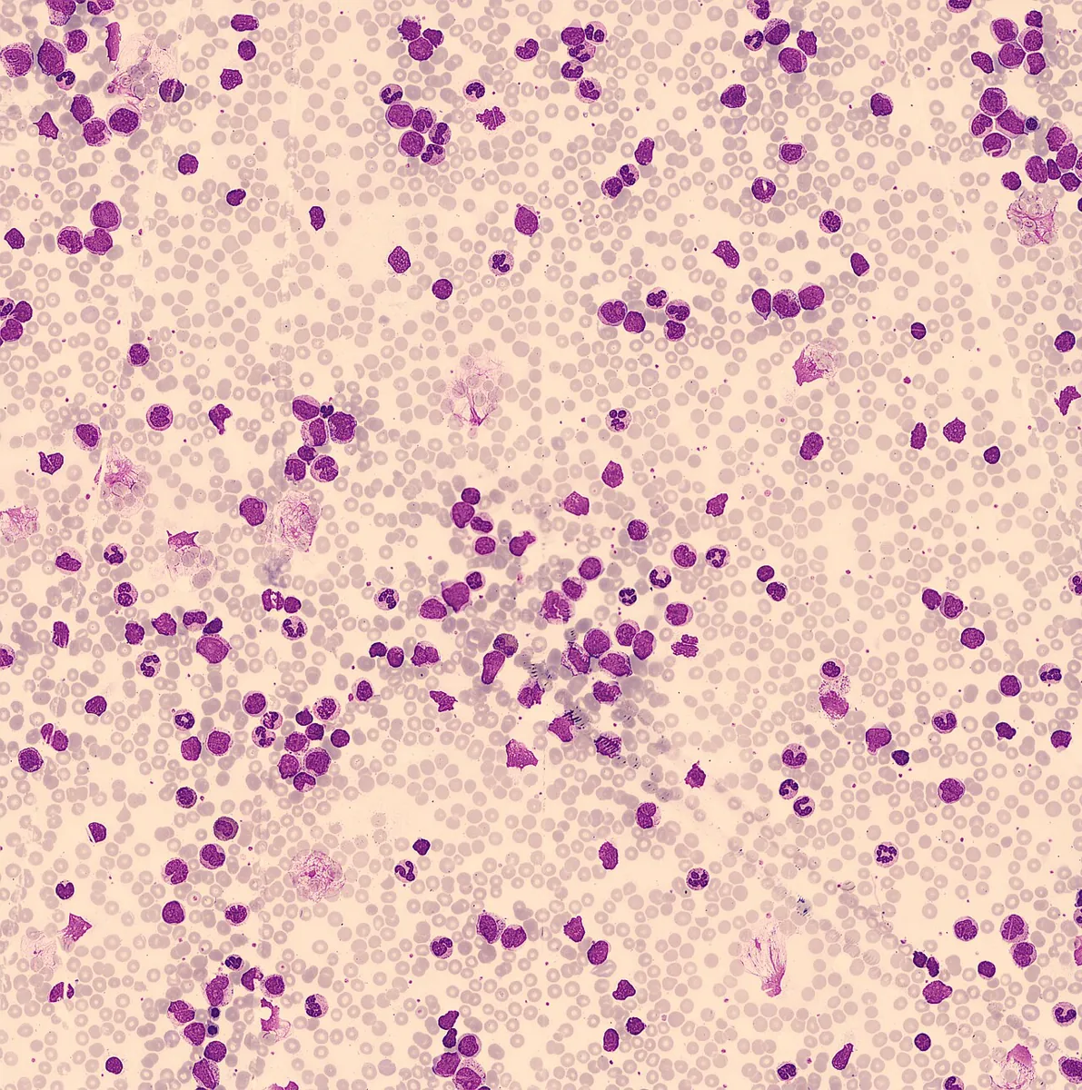
- Peripheral smear: immature granulocytes at all stages of maturation.
- Bone marrow: karyotype, cellularity, and extent of myelofibrosis.
- Detection of the Philadelphia chromosome or the BCR::ABL1 fusion by cytogenetics, FISH, or RT-PCR confirms the diagnosis; it is absent on classic cytogenetics in about 5 %.
- Advanced phase: anemia, thrombocytopenia, additional chromosomal abnormalities (e.g., trisomy 8, isochromosome 17q).
Keep learning in the app
Further reading (open access)
- MSD Manual Professional: Chronic Myeloid Leukemia (CML)
- Khoury et al., Leukemia 2022: WHO-Klassifikation (5. Aufl.), myeloische Neoplasien (PMC-Volltext)
- MSD Manual Professional: Overview of Leukemia
- Arber et al., Blood 2022: International Consensus Classification (ICC) myeloischer Neoplasien und akuter Leukämien (PMC-Volltext)
Cross-references
More topics: Haematology & oncology
- Anemia (classification and work-up)
- Hemolytic anemias
- Acute myeloid leukemia (AML)
- Iron deficiency anemia
- Non-Hodgkin lymphomas
- Vitamin B12 deficiency and pernicious anemia
- Multiple myeloma
- Paraneoplastic syndromes
- Autoimmune hemolytic anemia (AIHA)
- Febrile neutropenia
- Immune thrombocytopenia (ITP)
- Renal anemia
Note: Learning content for medical education – not a treatment recommendation and no substitute for diagnosis or treatment decisions in individual cases. Treatment and management are deliberately not covered on this page.