Chronic lymphocytic leukemia (CLL)
Board exam relevance: in 1 of 105 exam reports · rank 181- Synonyms
- chronic lymphatic leukemia, B-CLL, small lymphocytic lymphoma, SLL, blood cancer
- Specialty
- Internal medicine · Haematology & oncology
- Images
- Blood smear & cytology 1 · CT 1 · Histology 1 · Gross specimen 1
- Last updated
- 10/2026 · Dr. Pascal Bafteh
Contents
Images (4)
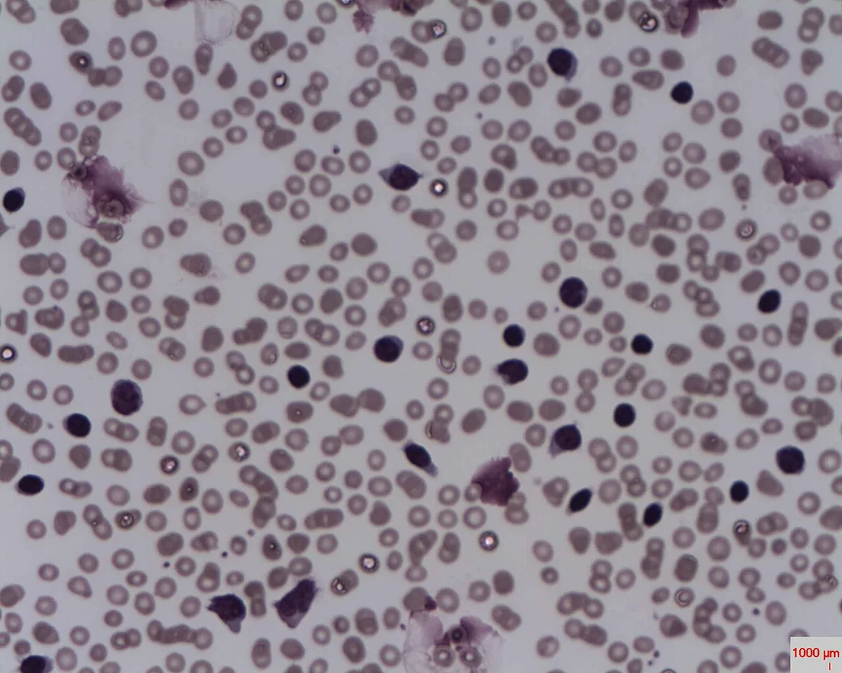 Blood smear & cytology
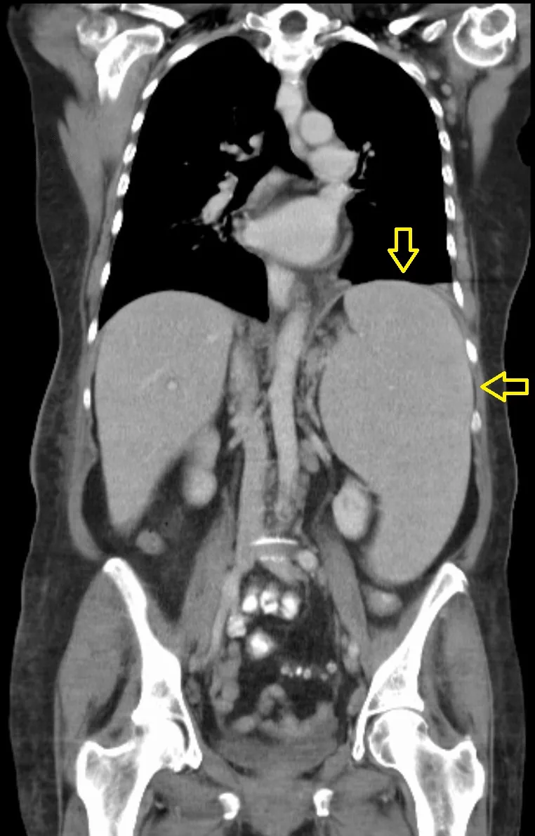 CT
CT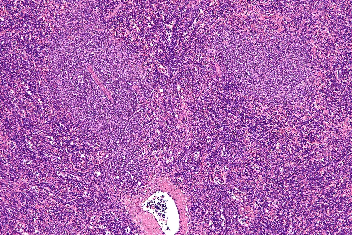 Histology
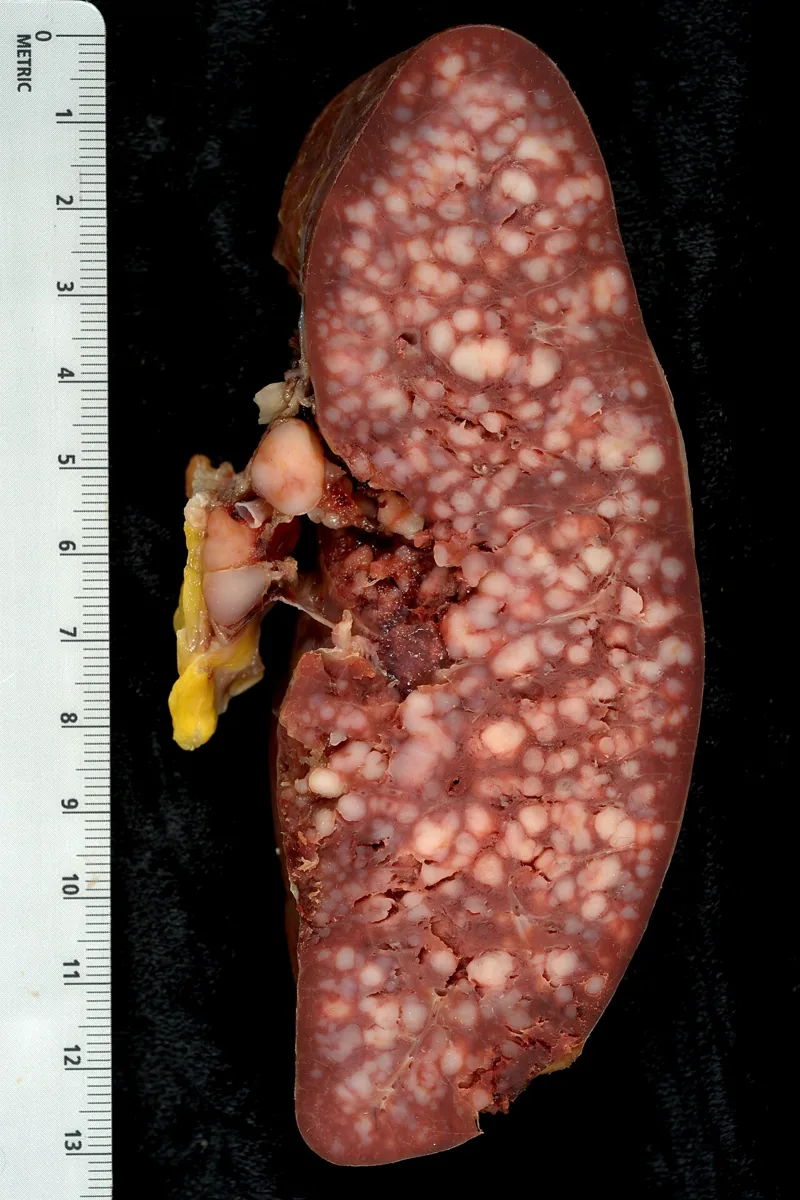 Gross specimen
Definition
Chronic lymphocytic leukemia (CLL) is a neoplasm of mature-appearing, monoclonal B lymphocytes that accumulate in the blood, bone marrow, lymph nodes, and spleen. CLL and small lymphocytic lymphoma (SLL) are regarded as one disease with different distribution. Its precursor is monoclonal B-cell lymphocytosis (MBL).
Classification
Binet stages (sites assessed: cervical, axillary, and inguinal lymph nodes, liver, spleen):
- Binet A: ≤ 2 involved sites, hemoglobin ≥ 10 g/dL, platelets ≥ 100,000/µL
- Binet B: 3–5 involved sites, hemoglobin and platelets as in stage A
- Binet C: hemoglobin < 10 g/dL or platelets < 100,000/µL, regardless of the number of involved sites
Rai stages: 0 lymphocytosis; I plus lymph node enlargement; II plus hepatomegaly or splenomegaly; III plus anemia (hemoglobin < 11 g/dL); IV plus thrombocytopenia (< 100,000/µL).
MBL according to WHO 2022: clonal B cells < 0.5 × 10⁹/L (low-count MBL) or ≥ 0.5 × 10⁹/L with total B cells < 5 × 10⁹/L (CLL/SLL-type MBL), each without other features of CLL.
Occurrence & epidemiology
CLL is the most common type of leukemia in the Western world. In Germany, according to the RKI, a good third of newly diagnosed leukemias are CLL. The average age at diagnosis is about 70 years; the disease is extremely rare in children. It is rare in Japan and China and more common in people of Ashkenazi Jewish ancestry.
Aetiopathogenesis
CD5-positive B cells acquire mutations, become continuously activated, and first form MBL; further genetic abnormalities lead to CLL. About 1–2 % of MBL progresses to CLL per year. A hereditary component is likely in some cases. Recurrent genetic abnormalities include del(13q), del(11q), del(17p), and trisomy 12, as well as TP53 mutations.
Clinical features
- Often asymptomatic initially; diagnosis frequently follows an incidental finding of lymphocytosis.
- Nonspecific complaints: fatigue, weakness, anorexia, weight loss, fever, night sweats, early satiety.
- Lymphadenopathy in more than 50 %, localized (mostly cervical and supraclavicular) or generalized; splenomegaly and hepatomegaly less common.
- Susceptibility to infection due to hypogammaglobulinemia, which develops in up to two-thirds.
- Autoimmune phenomena: autoimmune hemolytic anemia (positive direct antiglobulin test) and immune thrombocytopenia.
- Richter transformation into diffuse large B-cell lymphoma in about 2–10 %.
Diagnosis
- Blood count: absolute lymphocytosis above 5,000/µL raises suspicion of CLL.
- Flow cytometry of blood: demonstrates clonality; coexpression of CD5, CD19, CD20, and CD23 with light chain restriction (kappa or lambda) is typical.
- Bone marrow examination is not required for diagnosis; if performed, often more than 30 % lymphocytes.
- Other findings: hypogammaglobulinemia, elevated LDH, uric acid, and liver enzymes, rarely hypercalcemia.
- Cytogenetics and molecular genetics from blood (e.g., FISH for del(17p), TP53 mutation, IGHV mutation status) for risk assessment.
Keep learning in the app
Further reading (open access)
Cross-references
More topics: Haematology & oncology
- Anemia (classification and work-up)
- Hemolytic anemias
- Acute myeloid leukemia (AML)
- Iron deficiency anemia
- Non-Hodgkin lymphomas
- Vitamin B12 deficiency and pernicious anemia
- Multiple myeloma
- Paraneoplastic syndromes
- Autoimmune hemolytic anemia (AIHA)
- Febrile neutropenia
- Immune thrombocytopenia (ITP)
- Renal anemia
Note: Learning content for medical education – not a treatment recommendation and no substitute for diagnosis or treatment decisions in individual cases. Treatment and management are deliberately not covered on this page.