Trichomoniasis
Exam relevance: in 4 of 197 board exam reports · rank 111
- Synonyms
- trich, Trichomonas vaginalis
- Specialty
- Gynaecology · Vulva & vagina
- Images
- Histology & cytology 2 · Colposcopy 1
- Exam relevance
- 4 of 197 reports · rank 111
- In the app
- 1 flashcards · GynFuchs
- Last updated
- 10/2026 · Dr. Pascal Bafteh
Contents
Images (3)
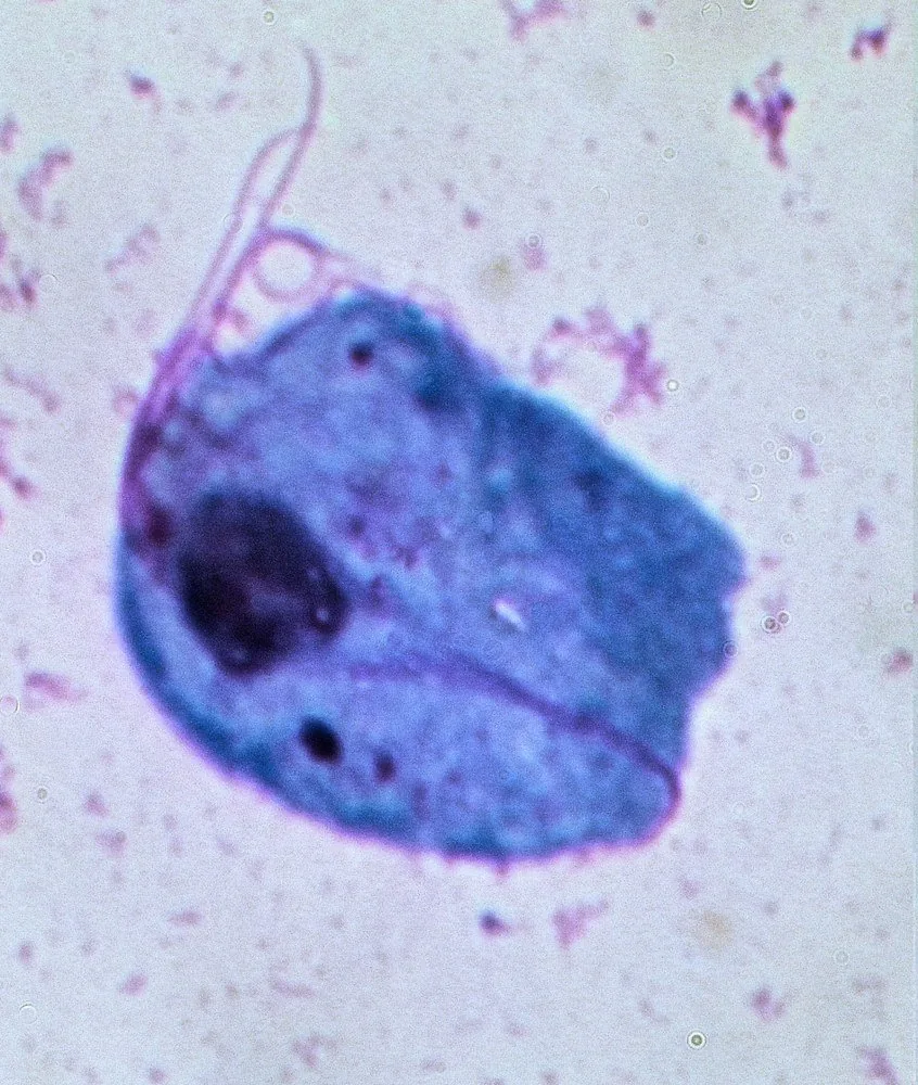 Histology & cytology
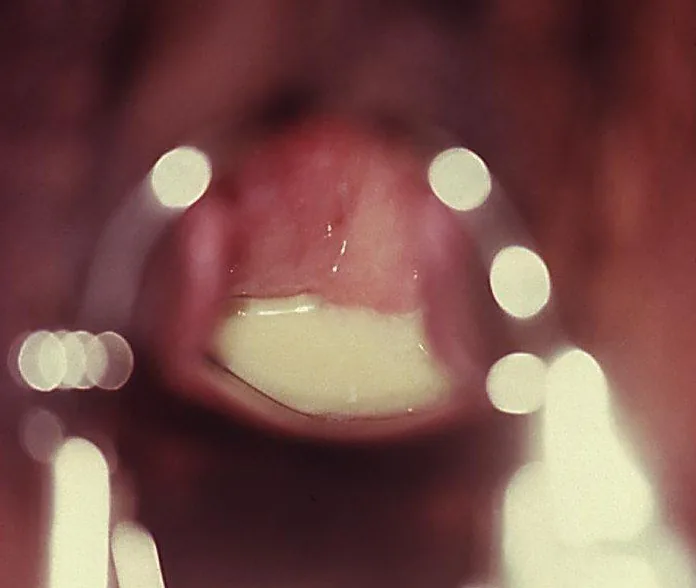 Colposcopy
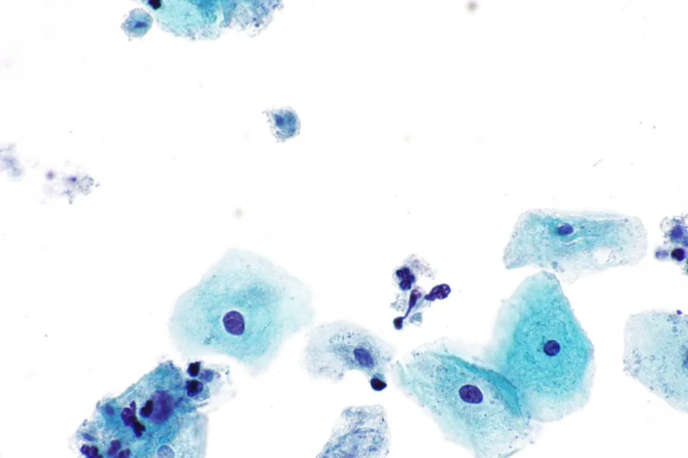 Histology & cytology
Definition
Pathogen and Clinical Presentation
- The pathogen is: Trichomonas vaginalis, a protozoan.
- Route of transmission: sexual — it is a sexually transmitted infection.
- Characteristic discharge: yellow-green, frothy, malodorous.
- The pH is: elevated (> 4.5).
- The classic but rare finding on the cervix: colpitis macularis (strawberry cervix).
- Trichomoniasis is a sexually transmitted infection with the unicellular, motile protozoan Trichomonas vaginalis, which lives in the lower genitourinary tract in women.
Occurrence & epidemiology
Epidemiology
- It is the most common non-viral sexually transmitted infection worldwide; nearly 156 million new infections were estimated for 2020.
- Women are affected more often than men; global prevalence in 2016 was estimated at 5.3 % in women and 0.6 % in men.
Aetiopathogenesis
Aetiology and risk factors
- Transmission is sexual; asymptomatically infected people, particularly men, can also pass on the organism over long periods.
- The infection is accompanied by an imbalance of the vaginal flora, a pro-inflammatory immune response and a raised vaginal pH.
- The infection increases the risk of transmission and acquisition of HIV and other sexually transmitted infections.
Clinical features
- A proportion of infections are asymptomatic but may become symptomatic at any time; coinfection with other sexually transmitted pathogens is common.
- Common complaints are discharge, itching, vulvar redness and swelling, dysuria and dyspareunia; a yellow-green, frothy, malodorous discharge is suggestive but rather unusual.
- In pregnancy, the infection is associated with low birth weight, preterm labour and early rupture of membranes.
Diagnosis
- Quickest method of detection: wet mount: motile, pear-shaped organisms.
- What spoils the diagnosis: cooled or dried-out specimen.
- Most reliable method of detection: nucleic acid test (PCR) from vaginal swab.
- Incidental finding possible: on Pap smear (as small grey cells).
Typical imaging findings
- Colposcopy: Abundant yellowish, thin and frothy discharge pools in the posterior fornix, running out of the os. The mucosa is reddened; pH is clearly alkaline.
- Punctate mucosal haemorrhages on the vaginal wall and cervix (strawberry cervix) are classic and visible on colposcopy in about 45 % of patients; vaginal pH is raised.
- Motile, pear-shaped, flagellated trichomonads on wet mount confirm the diagnosis; sensitivity varies widely, as the organisms can become immotile within minutes of slide preparation.
- Nucleic acid amplification tests (NAAT) are more sensitive than microscopy or culture and have largely replaced them as the reference method.
Keep learning in the app
Further reading (selection)
Cross-references
Note: Learning content from the GynFuchs app (flashcards, exam questions, image cases) for medical education – not a treatment recommendation and no substitute for diagnosis or treatment decisions in individual cases. Treatment and management are deliberately not covered on this page.