Vulvar melanoma
- Specialty
- Gynaecology · Vulva & vagina
- Images
- Histology & cytology 1 · Clinical 1
- In the app
- 1 flashcards · GynFuchs
- Last updated
- 10/2026 · Dr. Pascal Bafteh
Contents
Images (2)
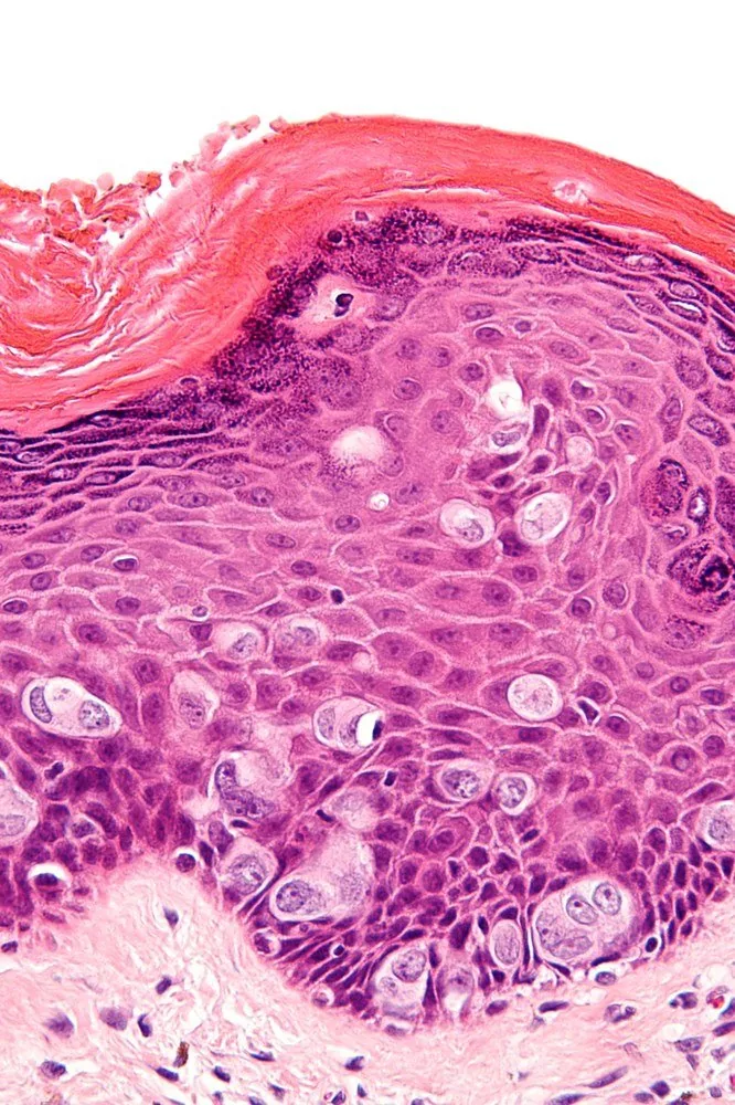 Histology & cytology
Definition
Vulvar Melanoma
- Incidence: Vulvar melanoma is the second most common malignant tumour of the vulva.
- Clinical presentation: Suspicious findings are newly appeared, irregularly pigmented, asymmetrical lesions with an indistinct border, bleeding, or an increase in size.
- Location: The most common locations are the clitoral region and the labia minora, i.e., non-hair-bearing parts.
- Prognostic factor: The decisive prognostic factor is the tumour thickness according to Breslow.
- Vulvar melanoma is a malignant melanocytic tumour of the vulva; the vulva is the most common site of melanoma in the female genital tract.
Classification
- The mucosal lentiginous subtype is the most common histologically; nodular and superficial spreading melanomas are also described.
Occurrence & epidemiology
Epidemiology
- It is the second most common malignant tumour of the vulva after squamous cell carcinoma.
- Mainly white women from the fifth to the seventh or eighth decade are affected; the mean age at diagnosis is 60–63 years.
Aetiopathogenesis
Aetiology and risk factors
- The relationship with sun exposure is undefined.
- KIT mutations are molecularly characteristic; BRAF and NRAS mutations also occur.
Clinical features
- Tumours occur, in descending order of frequency, on the labia majora, labia minora and clitoral hood, mainly on hairless mucosa; only about 13 % affect the outer hair-bearing skin.
- Symptoms are non-specific: itching, bleeding or atypical discharge and a sensation of a lump, with lymphadenopathy in advanced disease; some tumours are asymptomatic.
- Amelanotic forms account for 2–10 % and are more common after the menopause; clinical presentation is often delayed because early symptoms are absent.
Diagnosis
- The diagnosis is made by biopsy and confirmed by immunohistochemistry, for amelanotic lesions for example with HMB-45, S-100, MART-1 and tyrosinase.
- Micro-staging follows Clark, Breslow and Chung; dermoscopy and confocal microscopy support the clinical assessment.
Keep learning in the app
Further reading (selection)
Cross-references
Note: Learning content from the GynFuchs app (flashcards, exam questions, image cases) for medical education – not a treatment recommendation and no substitute for diagnosis or treatment decisions in individual cases. Treatment and management are deliberately not covered on this page.