Vulvar cancer
Exam relevance: in 8 of 197 board exam reports · rank 82
- Synonyms
- vulval cancer
- Specialty
- Gynaecology · Vulva & vagina
- Images
- Clinical 3
- Exam relevance
- 8 of 197 reports · rank 82
- In the app
- 2 flashcards · GynFuchs
- Last updated
- 10/2026 · Dr. Pascal Bafteh
Contents
Images (3)
Classification
Lymphatic Drainage
- Drainage pathways: The lymphatic drainage of the vulva proceeds first to the superficial and deep inguinofemoral lymph nodes, then onwards to the pelvic nodes.
- Lateral tumours: For laterally located tumours with a distance of at least 1 cm from the midline, ipsilateral assessment is sufficient.
Staging
- FIGO I: Stage FIGO I means a tumour confined to the vulva or perineum without lymph node metastases.
- FIGO IA: Stage FIGO IA is defined as a tumour up to 2 cm with a depth of invasion up to 1 mm.
- FIGO II: Stage FIGO II means spread to the lower parts of the urethra, vagina, or anus without lymph node metastases.
- FIGO III: Stage FIGO III means lymph node metastases in the inguinofemoral lymph nodes.
- FIGO IV: Stage FIGO IV means involvement of upper urethral or vaginal parts, bladder or rectal mucosa, pelvic bone, fixed or ulcerated inguinal lymph nodes, or distant metastases.
Occurrence & epidemiology
Epidemiology and Aetiology
- Histology: The most common histological type of vulvar carcinoma is squamous cell carcinoma.
- Pathways of development: Two main pathways of development are distinguished, namely the HPV-dependent pathway via HSIL in younger women and the HPV-independent pathway via lichen sclerosus and dVIN in older women.
- Incidence: The incidence of vulvar carcinoma has increased in recent decades, including in younger women.
Aetiopathogenesis
Risk Factors
- HPV-associated risks: HPV-associated risk factors include persistent infection with high-risk types, smoking, immunosuppression, and concurrent cervical neoplasia.
- HPV-independent risk: The most important HPV-independent risk factor is lichen sclerosus.
Clinical features
Clinical Presentation and Predilection Sites
- Further symptoms: Other complaints include a non-healing sore, bleeding, burning, pain, and a palpable lump.
- Predilection sites: Typical predilection sites are the labia majora, followed by the labia minora, clitoral region, and posterior commissure.
Histology
Other Histologies
- Rare entities: Other rare entities are basal cell carcinoma, adenocarcinoma of the Bartholin's gland, and sarcoma.
Diagnosis
Diagnosis and Staging
- Diagnostic confirmation: The diagnosis is confirmed by a punch biopsy from the edge of the lesion.
- Histopathology: Information from the biopsy regarding histological type, grading, depth of invasion, and the HPV or p16 and p53 status is crucial.
- Clinical examination: The clinical examination includes the entire vulva with vulvoscopy, vagina, and cervix, as well as the inguinal lymph nodes.
- Staging investigations: Staging includes ultrasound of the groins, and in advanced cases, cross-sectional imaging of the pelvis and abdomen, cystoscopy and rectoscopy as needed.
Typical imaging findings
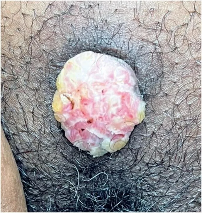
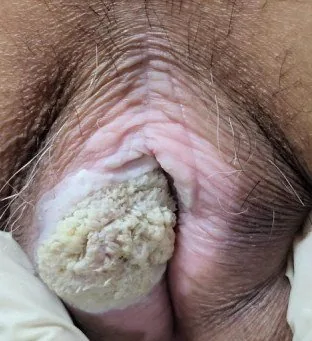
- Clinical finding: A rounded, firm, exophytic nodule with an irregular, partly keratotic and eroded surface lies on the labium. The margin is raised and the lesion is barely mobile against the underlying tissue; the surrounding skin appears atrophic.
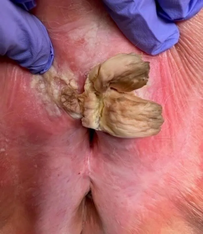
- Clinical finding: A firm, yellowish-brown horn-like structure protrudes from a reddened, thickened base. The base itself is raised and indurated; the surrounding skin is pale, atrophic and shows signs of a chronic underlying disease.
Keep learning in the app
Further reading (selection)
Cross-references
Note: Learning content from the GynFuchs app (flashcards, exam questions, image cases) for medical education – not a treatment recommendation and no substitute for diagnosis or treatment decisions in individual cases. Treatment and management are deliberately not covered on this page.