Vulvar haematoma
- Specialty
- Gynaecology · Vulva & vagina
- Images
- Clinical 1
- Last updated
- 10/2026 · Dr. Pascal Bafteh
Contents
Images (1)
Definition
- A vulvar haematoma is a collection of blood in the vulva, which is supplied mainly by the pudendal artery.
- Injury to the labial branches of the internal pudendal artery readily initiates a haematoma; the source of bleeding is often a branch of the pudendal artery.
Occurrence & epidemiology
Epidemiology
- In obstetrics, vulvar haematoma is a recognised complication, although reported frequencies vary widely; outside obstetrics it is rare.
Aetiopathogenesis
Aetiology and risk factors
- During labour, they result from direct injury such as birth lacerations, episiotomy or instrumental vaginal birth, or indirectly from extensive stretching of the birth canal.
- Risk factors include hypertension, multiple pregnancy, a large baby, clotting abnormalities, episiotomy, vulvovaginal varicosities and first pregnancy; spontaneous haematomas typically follow precipitate labour or a prolonged second stage.
- Outside obstetrics, they mostly result from trauma such as straddle injuries or assault; rarely they occur after intercourse or spontaneously from rupture of the iliac or pudendal artery.
Clinical features
- The most common leading symptom is pain, after birth usually within 24 hours, together with vulvar swelling.
- After birth, the haematoma is detected at a median of about 6 hours; rarely it becomes apparent only days later.
- Possible consequences are voiding difficulties up to urinary retention, anaemia and haemodynamically significant haemorrhage, and tissue necrosis.
Diagnosis
Typical imaging findings
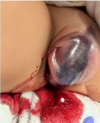
- Clinical finding: One labium is distended into a tensely swollen, blue-livid mass that distorts the vulva asymmetrically. The overlying skin is shiny and taut; fresh blood is seen at the perineum.
- Clinically, there is a tender, fluctuant swelling, because bleeding into the vulva is largely restricted only by Colles fascia and the urogenital diaphragm.
- Ultrasound helps to distinguish it from an abscess or cyst, and CT angiography can show the bleeding vessel and active extravasation; there is no gold standard for imaging.
Keep learning in the app
Further reading (selection)
- Delayed presentation of a non-resorbing postpartum vulvar hematoma: A case report (Case Rep Womens Health 2023, PubMed Central)
- Computed Tomography Findings in Non-Obstetric Vulvar Hematoma: A Case Report (J Educ Teach Emerg Med 2024, PubMed Central)
- Postpartum spontaneous vulvar hematoma as a cause of maternal near miss: a case report and review of the literature (J Med Case Rep 2022, PubMed Central)
Cross-references
Note: Learning content from the GynFuchs app (flashcards, exam questions, image cases) for medical education – not a treatment recommendation and no substitute for diagnosis or treatment decisions in individual cases. Treatment and management are deliberately not covered on this page.