Vulvar intraepithelial neoplasia (VIN)
Exam relevance: in 6 of 197 board exam reports · rank 93
- Specialty
- Gynaecology · Vulva & vagina
- Images
- Histology & cytology 2
- Exam relevance
- 6 of 197 reports · rank 93
- In the app
- 1 flashcards · GynFuchs
- Last updated
- 10/2026 · Dr. Pascal Bafteh
Contents
Images (2)
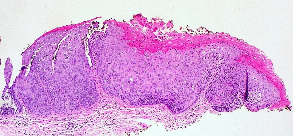 Histology & cytology
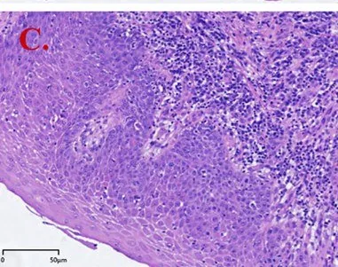 Histology & cytology
Classification
Two Pathways
- Nomenclature: HPV-dependent VIN is now referred to as vulvar HSIL or uVIN.
- uVIN characteristics: It affects younger women, is often multifocal, and is associated with smoking and immunosuppression.
- uVIN immunohistochemistry: Immunohistochemically, it is p16-positive as an expression of HPV transformation.
- HPV-independent form: The HPV-independent form is called differentiated VIN or dVIN.
- dVIN characteristics: It usually develops on a background of lichen sclerosus in older women and is mostly unifocal.
- dVIN immunohistochemistry: It is often p53-aberrant and p16-negative.
Why the Distinction Matters
- Diagnostics: It is also more difficult to recognise because it appears histologically mature and atypia is confined to the basal layer.
Clinical features
Clinical Presentation and Diagnosis
- Clinical picture: Clinically, VIN presents as a white, reddish, or pigmented lesion, raised or flat, often with pruritus.
- Confirming the diagnosis: The diagnosis is established by biopsy, or mapping with multiple punch biopsies in case of multifocal findings.
- Additional workup: An additional workup is mandatory for the vagina, cervix, and anal canal, as HPV-associated neoplasia can be multicentric.
Keep learning in the app
Further reading (selection)
Cross-references
Note: Learning content from the GynFuchs app (flashcards, exam questions, image cases) for medical education – not a treatment recommendation and no substitute for diagnosis or treatment decisions in individual cases. Treatment and management are deliberately not covered on this page.