Placental insufficiency
Exam relevance: in 2 of 197 board exam reports · rank 144
- Specialty
- Obstetrics · Pregnancy disorders
- Images
- Ultrasound 2
- Exam relevance
- 2 of 197 reports · rank 144
- In the app
- 1 flashcards · GynFuchs
- Last updated
- 10/2026 · Dr. Pascal Bafteh
Contents
Images (2)
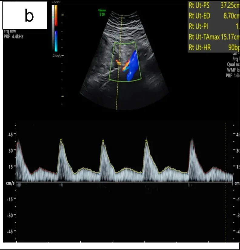 Ultrasound
 Ultrasound
Definition
- Placental insufficiency denotes inadequate placental perfusion that leads to an insufficient supply of oxygen and nutrients to the fetus; there is no universally accepted definition.
- It is understood as a progressive decline in placental function culminating in chronic fetal hypoxaemia and growth restriction.
Classification
- By time of diagnosis, early-onset (before 32 weeks) and late-onset growth restriction (from 32 weeks) are distinguished; the early form is usually more severe and often associated with pre-eclampsia.
Aetiopathogenesis
Physiology and pathophysiology
- Doppler sonography of the uterine arteries reflects the maternal side of placentation, particularly the remodeling of spiral arteries.
- Physiologically, there is a significant drop in resistance until about 24th week of gestation.
- Pathological findings include a pulsatility index (PI) above the 95th percentile and a persistent notch, an incisure post-systolic.
Clinical features
Clinical features and complications
- Placental insufficiency contributes to pre-eclampsia, fetal growth restriction, oligohydramnios and stillbirth.
- As an adaptation to hypoxaemia, blood flow is redistributed to vital organs (brain sparing), fetal movements decrease and growth slows.
Histology
- Typical histopathological findings are distal villous hypoplasia, increased syncytial knots, maternal vascular malperfusion, placental infarcts and fibrin deposition; there is no pathognomonic marker.
Diagnosis
Typical imaging findings
- Ultrasound: The waveform rises steeply and then falls, producing a clear incisure immediately after systole; only then does diastolic flow continue at a low level. The pattern repeats identically with every heartbeat.
- Classically, growth restriction is defined by an estimated fetal weight or abdominal circumference below the 10th centile; fetal mortality is highest below the 3rd centile.
- Diagnosis relies mainly on ultrasound biometry and Doppler velocimetry, especially of the umbilical artery.
Keep learning in the app
Further reading (selection)
Cross-references
Note: Learning content from the GynFuchs app (flashcards, exam questions, image cases) for medical education – not a treatment recommendation and no substitute for diagnosis or treatment decisions in individual cases. Treatment and management are deliberately not covered on this page.