Pre-eclampsia
Exam relevance: in 42 of 197 board exam reports · rank 9
- Synonyms
- preeclampsia, toxaemia of pregnancy
- Specialty
- Obstetrics · Pregnancy disorders
- Images
- Ultrasound 5 · Clinical 1
- Exam relevance
- 42 of 197 reports · rank 9
- In the app
- 4 flashcards · GynFuchs
- Last updated
- 10/2026 · Dr. Pascal Bafteh
Contents
Images (6)
 Ultrasound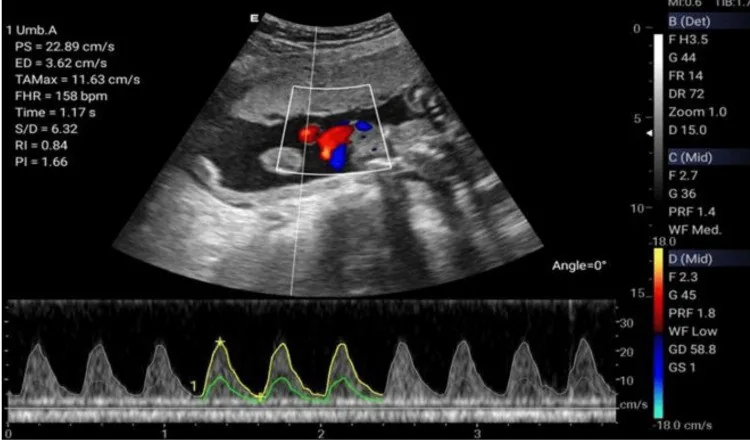
 Ultrasound
 Ultrasound
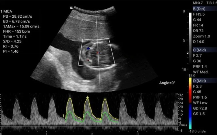 Ultrasound
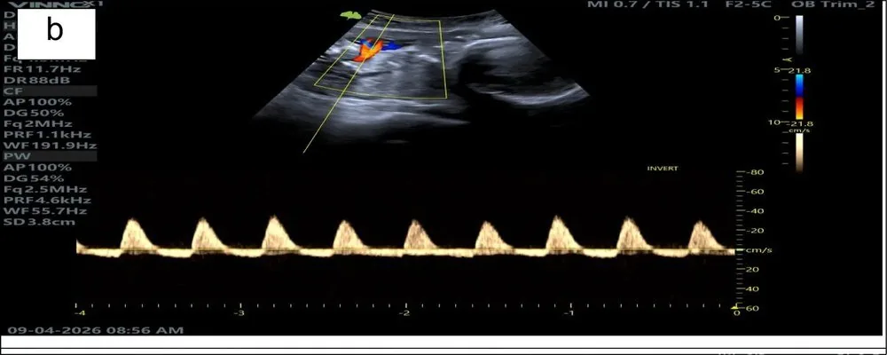 Ultrasound
Definition
Definition and diagnostic criteria
- Pre-eclampsia is defined by a new onset of blood pressure of ≥ 140/90 mmHg after 20+0 weeks.
Classification
Types and risk factors
- Early-onset pre-eclampsia (before 34+0 weeks) is typically placental with growth restriction.
- The single strongest risk factor for developing pre-eclampsia is history of pre-eclampsia.
- Other major risk factors include chronic hypertension, kidney disease, diabetes mellitus, and autoimmune diseases (SLE, APS).
Pre-eclampsia and superimposed pre-eclampsia
- Pre-eclampsia is present when gestational hypertension is accompanied by proteinuria or at least one other maternal organ manifestation or uteroplacental dysfunction.
- The former classification into 'mild' and 'severe' pre-eclampsia has been abandoned; instead, today the organ manifestations are named.
- If pre-eclampsia develops on the basis of pre-existing hypertension, it is called superimposed pre-eclampsia.
Special forms and diagnostics
- HELLP syndrome and eclampsia are considered severe manifestation and not separate primary categories.
Aetiopathogenesis
Impaired placentation
- The two-stage model begins with an incomplete invasion of the trophoblast into the spiral arteries.
- Consequently, the physiological remodelling into wide, low-resistance vessels incomplete.
- The consequence of this first stage is placental malperfusion with oxidative stress, which initiates the second stage of the disease.
Anti-angiogenesis and endothelial damage
- The ischaemic placenta releases increased levels of anti-angiogenic factors, primarily sFlt-1 (soluble fms-like tyrosine kinase-1) and endoglin.
- sFlt-1 leads to a deficiency of free pro-angiogenic factors by binding and inactivating PlGF and VEGF.
- The clinical result is a generalised endothelial dysfunction with hypertension, oedema and proteinuria.
Clinical features
Clinical organ involvement
- Liver involvement presents with right-sided or epigastric upper abdominal pain, as well as transaminases that are are more than double the normal limit.
- Haematological signs include thrombocytopenia with counts < 100,000/µl, haemolysis, or disseminated intravascular coagulation.
- Neurological symptoms include new-onset headaches, visual disturbances such as flashing scotomas, or hyperreflexia and clonus.
- A uteroplacental manifestation can present as growth restriction/abnormal Doppler.
More facts from the study questions
- In the absence of proteinuria, a single other new-onset organ manifestation is sufficient for diagnosis.
- The diagnostic criteria require hypertension plus either proteinuria or other organ involvement.
- A history of pre-eclampsia in a previous pregnancy is the strongest single risk factor.
- Other risk factors include chronic hypertension, diabetes mellitus and autoimmune diseases.
- The threshold for relevant thrombocytopenia in pre-eclampsia is a count below 100,000 per µl.
- Counts between 100,000 and 150,000 per µl are low but do not define the haematological criterion.
- Liver involvement can present with right upper quadrant or epigastric pain.
- A rise in transaminases to more than double the normal value is a diagnostic criterion.
- The definition of severe hypertension in pregnancy is blood pressure values of 160/110 mmHg or higher.
- If a patient with pre-existing chronic hypertension newly develops the criteria for pre-eclampsia, this is termed superimposed pre-eclampsia.
Diagnosis
Diagnostic correlates
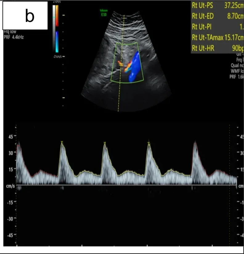
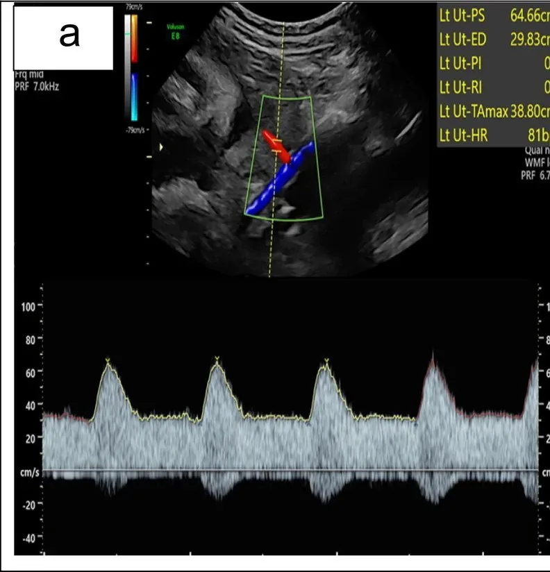
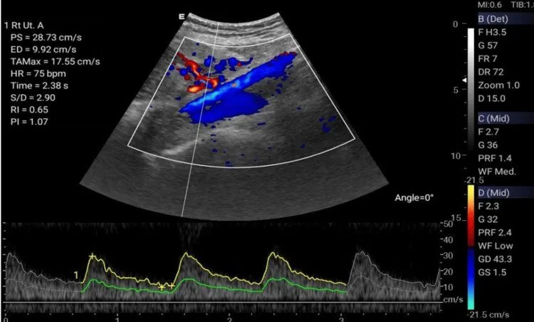
- A sonographic correlate of the impaired invasion in the first stage is an increased PI and notch in uterine arteries.
- The key laboratory correlate of the second stage is the elevated sFlt-1/PlGF ratio.
- A low sFlt-1/PlGF ratio rules out pre-eclampsia short-term.
History and clinical examination
- In addition to standardised blood pressure measurement and lung auscultation, the clinical examination includes assessing the reflex status with clonus testing.
- A rapid increase in oedema and weight is also an important important warning sign.
Laboratory and urine tests
- Urinalysis includes a dipstick test and measurement of the protein-to-creatinine ratio.
- The so-called HELLP panel includes a full blood count and specifically haptoglobin, LDH, as well as transaminases (AST, ALT).
- A specific marker for risk stratification is the serum sFlt-1/PlGF ratio.
Typical imaging findings
- Ultrasound: The waveform rises steeply and then falls, producing a clear incisure immediately after systole; only then does diastolic flow continue at a low level. The pattern repeats identically with every heartbeat.
Keep learning in the app
Further reading (selection)
Cross-references
Note: Learning content from the GynFuchs app (flashcards, exam questions, image cases) for medical education – not a treatment recommendation and no substitute for diagnosis or treatment decisions in individual cases. Treatment and management are deliberately not covered on this page.