Placenta praevia
Exam relevance: in 6 of 197 board exam reports · rank 93
- Synonyms
- placenta previa, low-lying placenta
- Specialty
- Obstetrics · Pregnancy disorders
- Images
- Ultrasound 2
- Exam relevance
- 6 of 197 reports · rank 93
- In the app
- 2 flashcards · GynFuchs
- Last updated
- 10/2026 · Dr. Pascal Bafteh
Contents
Images (2)
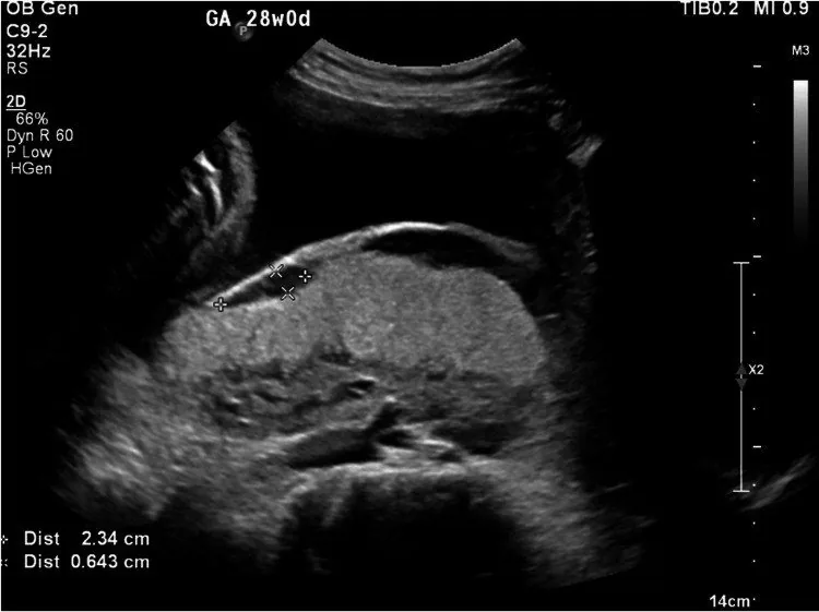 Ultrasound
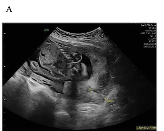 Ultrasound
Classification
- Classically: complete, partial and marginal placenta praevia, plus a low-lying placenta.
- The simplification used today is praevia when the internal os is covered, and low-lying when the edge is within 20 mm.
- The diagnosis is made transvaginally, which measures the distance to the internal os most accurately.
Clinical features
Risk factors and presentation
- Typical is sudden, painless, bright red bleeding, often waking her at night.
- The uterus is soft and not tender, and the CTG is usually normal at first.
Diagnosis
Measurement
- What is assessed is the wall (anterior, posterior, fundal, lateral) and the distance from the placental edge to the internal os.
- The most accurate is a transvaginal measurement with an empty bladder.
- A full bladder or a contraction of the lower segment can mimic praevia.
- The placental site belongs in the second screening scan of routine antenatal care.
"migration"
- A low-lying placenta in the second trimester resolves in the great majority of cases by the third.
- The reason is growth of the lower uterine segment, not true movement of the placenta.
- So a check is made in the third trimester, around 32 weeks.
- The further the placenta overlaps the os initially, the less likely it is to resolve.
What else is checked
- After a previous caesarean the relation of the placenta to the scar is assessed, because that is where the accreta risk lies.
- With a low-lying placenta you also look for vasa praevia with colour Doppler.
Typical imaging findings
- Ultrasound: The placenta lies on the posterior wall and extends across the internal os, which it completely covers. No free distance between placenta and cervix can be measured; the myometrium beneath the placenta is preserved.
Keep learning in the app
Further reading (selection)
Cross-references
Note: Learning content from the GynFuchs app (flashcards, exam questions, image cases) for medical education – not a treatment recommendation and no substitute for diagnosis or treatment decisions in individual cases. Treatment and management are deliberately not covered on this page.