Placenta accreta spectrum
Exam relevance: in 4 of 197 board exam reports · rank 111
- Specialty
- Obstetrics · Pregnancy disorders
- Images
- Ultrasound 1 · Clinical 1
- Exam relevance
- 4 of 197 reports · rank 111
- In the app
- 1 flashcards · GynFuchs
- Last updated
- 10/2026 · Dr. Pascal Bafteh
Contents
Images (2)
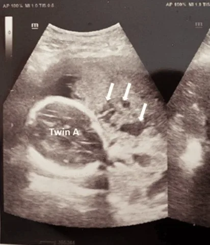 Ultrasound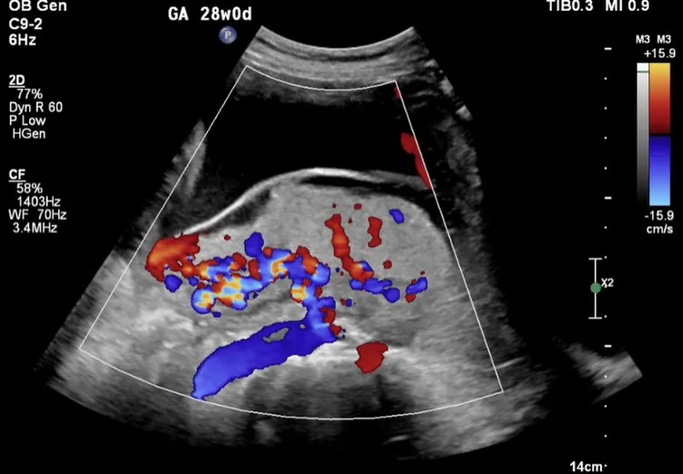
Definition
- Placenta accreta spectrum comprises disorders of placentation with abnormal invasion of trophoblast into the myometrium.
Classification
Terms
- Placenta accreta means villi attaching directly to the myometrium because decidua is missing.
- Increta means growth into the myometrium, and percreta growth through the serosa, possibly involving bladder or parametria.
- The strongest risk factor is the combination of placenta praevia and previous caesarean, rising with the number of caesareans.
- By depth of invasion, the following are distinguished: placenta accreta (villi attach directly to the myometrium in the absence of decidua), increta (villi penetrate deeply into the myometrium) and percreta (villi reach or penetrate the serosa).
- FIGO has proposed a grading system under the umbrella term PAS that replaces the previous categorical terminology.
Occurrence & epidemiology
Epidemiology
- Its frequency has increased with the rising caesarean section rate; a US database study found placenta accreta in 1 in 272 births between 1998 and 2011.
Aetiopathogenesis
Aetiology and risk factors
- The main risk factor is previous caesarean section combined with placenta praevia; with placenta praevia the risk rises with the number of caesarean sections from 3% (none) through 11%, 40% and 61% to 67% (four or more).
Clinical features
Clinical features and complications
- PAS is associated with increased maternal morbidity and mortality; after birth of the baby the placenta often does not separate within 30 minutes, and attempts at separation typically cause massive haemorrhage.
Diagnosis
Ultrasound signs
- Typical are loss of the retroplacental clear zone, placental lacunae ("Swiss cheese"), thinning of the myometrium and interruption of the bladder wall line.
- On colour Doppler you see hypervascularity at the uterovesical interface and bridging vessels between placenta and surroundings.
- MRI is considered for a posterior placenta, unclear ultrasound findings and assessment of the extent.
- MRI has high sensitivity and specificity but has not so far proved superior to ultrasound; it is used mainly when ultrasound findings are inconclusive.
Keep learning in the app
Further reading (selection)
Cross-references
Note: Learning content from the GynFuchs app (flashcards, exam questions, image cases) for medical education – not a treatment recommendation and no substitute for diagnosis or treatment decisions in individual cases. Treatment and management are deliberately not covered on this page.