Parvovirus B19 in pregnancy
Exam relevance: in 10 of 197 board exam reports · rank 66
- Synonyms
- fifth disease, slapped cheek
- Specialty
- Obstetrics · Pregnancy disorders
- Images
- Ultrasound 1
- Exam relevance
- 10 of 197 reports · rank 66
- In the app
- 1 flashcards · GynFuchs
- Last updated
- 10/2026 · Dr. Pascal Bafteh
Contents
Images (1)
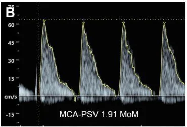 Ultrasound
Definition
Pathogen and Clinical Presentation
- In children, the disease is called: fifth disease, i.e., erythema infectiosum.
- Typical in children: malar erythema, lacy exanthem.
- What the virus attacks: erythroid progenitor cells.
- The fetus is therefore at risk of: aplastic anaemia, hydrops fetalis.
- Predominant in adults: arthralgia, flu-like course.
- The child is contagious: before the exanthem appears.
- Parvovirus B19 infection in pregnancy is a maternal infection with the agent of erythema infectiosum (fifth disease) that can be transmitted transplacentally to the fetus.
Occurrence & epidemiology
Epidemiology
- At the start of pregnancy, 34–65% of pregnant women are not immune, depending on the population studied.
Aetiopathogenesis
Aetiology and pathogenesis
- The virus is transmitted via respiratory secretions, hand-to-mouth contact, blood products and transplacentally.
- Fetal tissues, including haematopoietic cells and myocardium, express the viral P antigen receptor, which mainly explains anaemia and hydrops.
- The rate of transmission to the fetus is 17–33%.
- The fetus is most at risk in the first and second trimesters; after 20 weeks of gestation the risk of hydrops is 1% or less.
Clinical features
Clinical features and complications
- In immunocompetent adults, infection is usually asymptomatic or mild; maternal manifestations include erythema infectiosum, arthropathy, anaemia and thrombocytopenia.
- Fetal consequences include anaemia, thrombocytopenia, myocarditis, effusions, non-immune hydrops and intrauterine death; however, most infected fetuses remain unaffected.
Diagnosis
Fetal Monitoring
- The crucial measurement is: PSV in the middle cerebral artery (MCA).
- What causes it to increase in anaemia: lower blood viscosity.
- Threshold for a suspected diagnosis of severe anaemia: PSV increase above 1.5 MoM.
- Other signs on ultrasound: ascites, effusions, skin & placental edema.
- Maternal diagnosis is based on IgG and IgM antibodies; IgM is detectable from the end of the first week of infection.
- Fetal infection is detected by PCR of amniotic fluid.
- A middle cerebral artery peak systolic velocity above 1.5 MoM is highly suggestive of moderate to severe fetal anaemia.
- Ultrasound signs of hydrops are ascites, skin oedema, pleural and pericardial effusions and placental oedema.
Keep learning in the app
Further reading (selection)
Cross-references
Note: Learning content from the GynFuchs app (flashcards, exam questions, image cases) for medical education – not a treatment recommendation and no substitute for diagnosis or treatment decisions in individual cases. Treatment and management are deliberately not covered on this page.