Adnexal torsion (ovarian torsion)
Exam relevance: in 11 of 197 board exam reports · rank 64
- Synonyms
- ovarian torsion, twisted ovary
- Specialty
- Gynaecology · Ovary & adnexa
- Images
- Ultrasound 3
- Exam relevance
- 11 of 197 reports · rank 64
- In the app
- 1 flashcards · GynFuchs
- Last updated
- 10/2026 · Dr. Pascal Bafteh
Contents
Images (3)
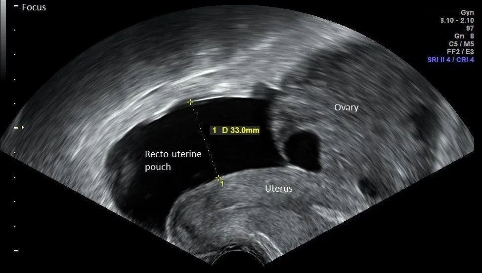 Ultrasound
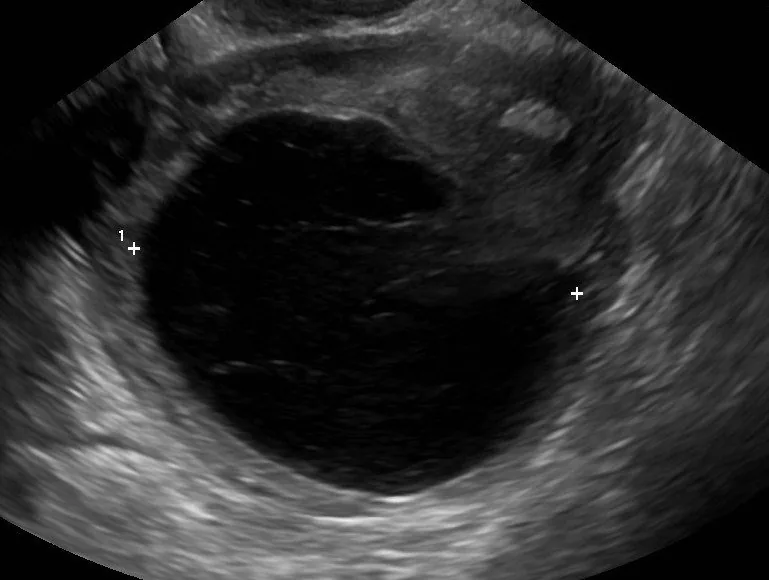 Ultrasound
 Ultrasound
Definition
Ovarian Torsion
- Symptomatology: Typical is sudden, severe, unilateral pain with nausea and vomiting, often wave-like.
- Ultrasound: On ultrasound, the ovary is enlarged and oedematous because arterial inflow is preserved longer than venous outflow.
- Doppler ultrasound: Doppler is of little help because preserved arterial flow does not exclude a torsion.
- In ovarian torsion the ovary twists on its supporting ligaments (infundibulopelvic and utero-ovarian ligaments); when the fallopian tube twists with it, the term adnexal torsion is used.
Aetiopathogenesis
Pathogenesis and risk factors
- First, venous and lymphatic outflow is compromised, with oedema, congestion and enlargement of the ovary; if torsion persists, ischaemia and haemorrhagic infarction follow.
- In women of reproductive age an underlying mass, usually larger than 5 cm, is present in 50–90%, most commonly a mature cystic teratoma.
- Endometriomas and malignancies cause torsion only occasionally because of fixation to adjacent structures.
Clinical features
- Typical is abrupt-onset lower abdominal pain radiating to the flank or groin with adnexal tenderness; often, however, the picture is non-specific with intermittent pain, low-grade fever, nausea and vomiting.
- Recurrent attacks may reflect episodes of torsion and detorsion; if unrecognised, necrosis, loss of the ovary and infertility may follow.
Diagnosis
- Ultrasound is the primary imaging method; because of the dual blood supply, colour Doppler may still show arterial flow, which does not exclude torsion.
- CT shows a unilaterally enlarged ovary, often displaced to the midline, with peripherally displaced follicles, a spirally twisted pedicle, free fluid and inflammatory fat stranding.
Keep learning in the app
Further reading (selection)
Cross-references
More topics: Ovary & adnexa
Note: Learning content from the GynFuchs app (flashcards, exam questions, image cases) for medical education – not a treatment recommendation and no substitute for diagnosis or treatment decisions in individual cases. Treatment and management are deliberately not covered on this page.