Hydrosalpinx
Exam relevance: in 1 of 197 board exam reports · rank 171
- Specialty
- Gynaecology · Ovary & adnexa
- Images
- Clinical 1 · Ultrasound 1
- Exam relevance
- 1 of 197 reports · rank 171
- Last updated
- 10/2026 · Dr. Pascal Bafteh
Contents
Images (2)
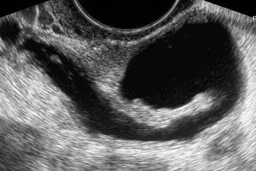
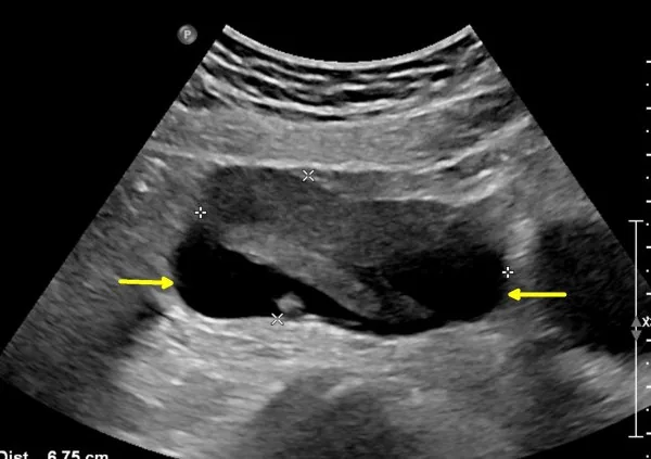 Ultrasound
Definition
- A hydrosalpinx develops when distal or proximal tubal occlusion causes the fallopian tube to fill with fluid and distend.
Occurrence & epidemiology
Epidemiology
- Tubal disease is involved in up to about 35% of cases of infertility.
- In a cohort after acute PID, 26.7% had a sonographic suspicion of hydrosalpinx after three months.
Aetiopathogenesis
Aetiology
- A hydrosalpinx is most often related to previous acute pelvic inflammatory disease, but it can also accompany a current acute salpingitis.
Clinical features
- In assisted reproduction, hydrosalpinx is associated with lower implantation and pregnancy rates and a higher risk of ectopic pregnancy and miscarriage.
- Proposed mechanisms include impaired endometrial receptivity and a toxic effect of the tubal fluid on early embryos.
Diagnosis
Typical imaging findings
- Ultrasound: Beside the ovary runs an elongated, anechoic, repeatedly folded cavity. Incomplete septa project from its wall into the lumen without dividing it fully; there are no solid parts.
- On ultrasound an elongated, convoluted cystic structure with anechoic or hypoechoic content and incomplete septa is typical; the septa are seen in up to 85–93% of chronic hydrosalpinges.
- Hyperechoic mural nodules of 2–3 mm (beads-on-a-string sign), which correspond to flattened, fibrotic remnants of the mucosal folds, indicate chronic hydrosalpinx.
- A thickened tubal wall and a cogwheel sign, by contrast, indicate acute salpingitis.
- In a meta-analysis of women with adnexal masses, transvaginal ultrasound reached a pooled sensitivity of 84% and a specificity of 99%.
- The gold standard is histological confirmation, although direct laparoscopic visualisation is also accepted.
- On CT and MRI hydrosalpinx appears as a fluid-filled, thin-walled tubular structure without wall enhancement, hyperintense on T2 and hypointense on T1 MRI.
Keep learning in the app
Further reading (selection)
Cross-references
More topics: Ovary & adnexa
- Ovarian cysts (adnexal cysts)
- Corpus luteum cyst and haemorrhagic ovarian cyst
- Mature cystic teratoma (dermoid cyst)
- Adnexal torsion (ovarian torsion)
- Ovarian cancer
- Borderline ovarian tumour
- Ovarian germ cell tumours
- Granulosa cell tumour and sex cord-stromal tumours
- Krukenberg tumour
- Pelvic inflammatory disease (PID)
Note: Learning content from the GynFuchs app (flashcards, exam questions, image cases) for medical education – not a treatment recommendation and no substitute for diagnosis or treatment decisions in individual cases. Treatment and management are deliberately not covered on this page.