Corpus luteum cyst and haemorrhagic ovarian cyst
Exam relevance: in 3 of 197 board exam reports · rank 125
- Specialty
- Gynaecology · Ovary & adnexa
- Images
- Ultrasound 4
- Exam relevance
- 3 of 197 reports · rank 125
- In the app
- 1 flashcards · GynFuchs
- Last updated
- 10/2026 · Dr. Pascal Bafteh
Contents
Images (4)
 Ultrasound
 Ultrasound
 Ultrasound
 Ultrasound
Definition
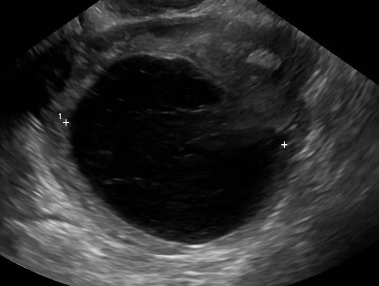
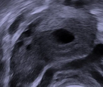
- When the corpus luteum fails to regress, a luteal cyst develops that may contain fluid or blood; the terms corpus luteum cyst and haemorrhagic corpus luteum are therefore often used synonymously.
Occurrence & epidemiology
Epidemiology
- In women of reproductive age most ovarian cysts are functional: dominant follicles, follicular cysts and luteal cysts.
- The corpus luteum is frequently seen on imaging in the second half of the cycle (luteal phase).
Aetiopathogenesis
Physiology
- After ovulation the dominant follicle collapses, and under the influence of LH the granulosa cells of the remaining follicular bed proliferate to form the corpus luteum.
- The corpus luteum produces mainly progesterone as well as inhibin A and oestradiol.
- In the absence of fertilisation its hormone secretion stops within 14 days of ovulation, and it regresses to a scar, the corpus albicans.
Clinical features
Cyst rupture and haemorrhage
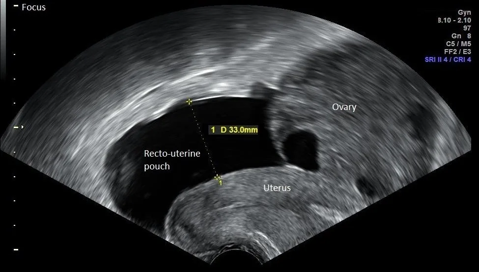
- Ultrasound finding: After a cyst rupture, one finds free fluid in the pouch of Douglas and an inhomogeneous, partially collapsed structure on the ovary.
- Rupture of a corpus luteum is the most common cause of gynaecological haemoperitoneum, with or without active bleeding.
Diagnosis
- On CT the corpus luteum appears as a well-circumscribed unilocular (rarely bilocular) cystic structure up to 3 cm with a crenulated, briskly enhancing rim, corresponding to the ring of fire sign on colour Doppler.
- A 3 cm threshold has been suggested: follicles and corpus luteum lie below it, functional follicular and luteal cysts above it; on CT both cyst types usually measure less than 5 cm.
- After intravenous contrast the walls of luteal cysts appear thicker and more vascularised than those of follicular cysts.
Keep learning in the app
Further reading (selection)
Cross-references
More topics: Ovary & adnexa
Note: Learning content from the GynFuchs app (flashcards, exam questions, image cases) for medical education – not a treatment recommendation and no substitute for diagnosis or treatment decisions in individual cases. Treatment and management are deliberately not covered on this page.