Ovarian germ cell tumours
Exam relevance: in 3 of 197 board exam reports · rank 125
- Specialty
- Gynaecology · Ovary & adnexa
- Images
- Histology & cytology 3
- Exam relevance
- 3 of 197 reports · rank 125
- In the app
- 2 flashcards · GynFuchs
- Last updated
- 10/2026 · Dr. Pascal Bafteh
Contents
Images (3)
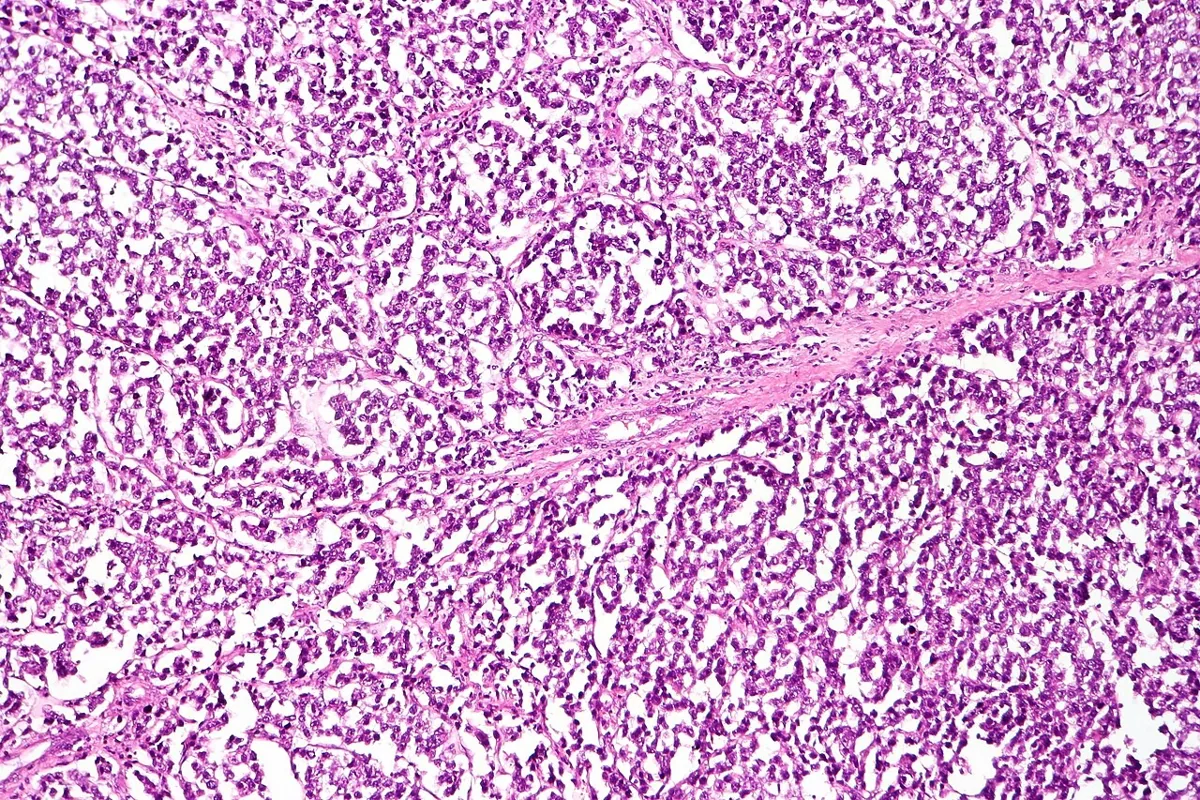 Histology & cytology
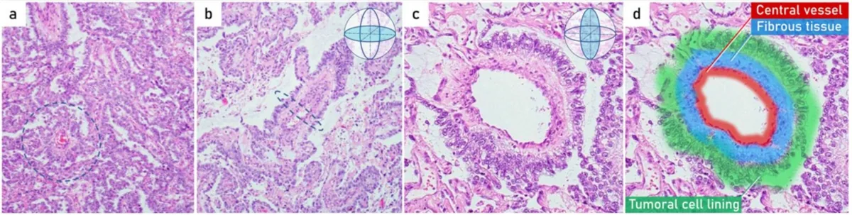 Histology & cytology
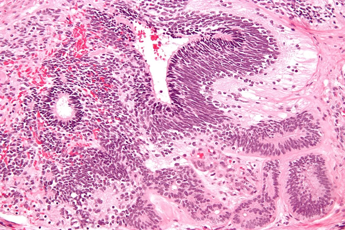 Histology & cytology
Definition
- Ovarian germ cell tumours arise from primitive germ cells; about 95% are benign mature cystic teratomas and only about 5% are malignant.
- Yolk sac tumour is a malignant primitive germ cell tumour that histologically resembles the mesenchyme of the primitive yolk sac.
Classification
Germ cell and sex cord-stromal tumours
- Most common germ cell tumour: The most common germ cell tumour is the mature teratoma, i.e., the dermoid cyst.
- Malignant germ cell tumours: Malignant germ cell tumours are dysgerminoma, yolk sac tumour, immature teratoma, and choriocarcinoma — typically in young women.
- Sex cord-stromal tumours: Sex cord-stromal tumours include granulosa cell tumour, theca cell tumour, fibroma, and Sertoli-Leydig cell tumour.
- Granulosa cell tumour: The granulosa cell tumour is clinically conspicuous due to oestrogen production with bleeding disorders, endometrial hyperplasia, or precocious puberty.
- Sertoli-Leydig cell tumour: The Sertoli-Leydig cell tumour causes virilisation due to androgen production.
Classification and epidemiology
- Among malignant types, in order of frequency, come dysgerminoma, immature teratoma, yolk sac tumour and mixed germ cell tumour; embryonal carcinoma, choriocarcinoma and malignant struma ovarii are rarer.
- Dysgerminoma accounts for about one third of malignant germ cell tumours; 80–85% of patients are under 30, and it is the most common ovarian tumour diagnosed in pregnancy and the puerperium.
- Malignant germ cell tumours are mostly unilateral, but pure dysgerminoma is bilateral in 10–15%.
Aetiopathogenesis
Aetiology
- Germ cell tumours can arise in dysgenetic gonads, for example in Turner or Swyer syndrome; with a Y chromosome a gonadoblastoma may develop, which undergoes malignant transformation in more than 40%.
Clinical features
- Occasionally the tumours produce hCG and cause signs of precocious puberty.
Histology
- Dysgerminomas are well encapsulated, firm and solid; tumour cells are surrounded by connective tissue containing lymphocytes, and diffuse strong nuclear OCT4 staining is characteristic.
- Schiller-Duval bodies are diagnostic of yolk sac tumour but are found in only about one third of cases.
Diagnosis
Tumour markers
- CA 19-9: The marker CA 19-9 is characteristically elevated in mucinous ovarian tumours and in pancreatic and biliary tract carcinomas.
- CEA: Carcinoembryonic antigen (CEA) is elevated primarily in gastrointestinal carcinomas, suggesting a metastasis in the case of an adnexal finding.
- Granulosa cell tumour: If a granulosa cell tumour is suspected, one measures inhibin B and anti-Müllerian hormone.
- Diagnosis relies on history, examination, ultrasound and the serum markers LDH, AFP and hCG: LDH is typical of dysgerminoma, AFP of yolk sac tumour and hCG of choriocarcinoma.
Keep learning in the app
Further reading (selection)
Cross-references
More topics: Ovary & adnexa
- Ovarian cysts (adnexal cysts)
- Corpus luteum cyst and haemorrhagic ovarian cyst
- Mature cystic teratoma (dermoid cyst)
- Adnexal torsion (ovarian torsion)
- Ovarian cancer
- Borderline ovarian tumour
- Granulosa cell tumour and sex cord-stromal tumours
- Krukenberg tumour
- Pelvic inflammatory disease (PID)
- Tubo-ovarian abscess
Note: Learning content from the GynFuchs app (flashcards, exam questions, image cases) for medical education – not a treatment recommendation and no substitute for diagnosis or treatment decisions in individual cases. Treatment and management are deliberately not covered on this page.