Ovarian cancer
Exam relevance: in 51 of 197 board exam reports · rank 6
- Specialty
- Gynaecology · Ovary & adnexa
- Images
- Endoscopy & gross 2 · Ultrasound 1
- Exam relevance
- 51 of 197 reports · rank 6
- In the app
- 4 flashcards · GynFuchs
- Last updated
- 10/2026 · Dr. Pascal Bafteh
Contents
Images (3)
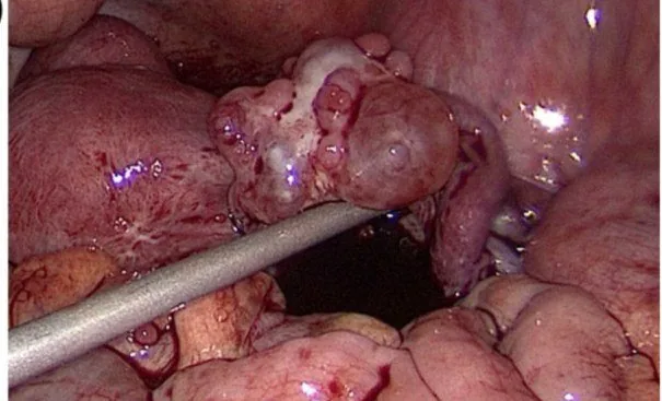 Endoscopy & gross
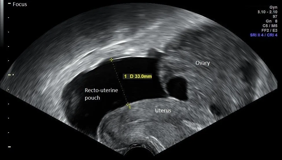 Ultrasound
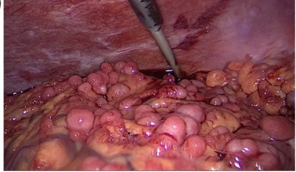 Endoscopy & gross
Classification
Stages
- FIGO I: FIGO stage I describes a tumour limited to the ovaries or fallopian tubes.
- FIGO IC: A stage FIGO IC is present with capsule rupture, tumour on the surface, or positive washings or ascites cytology.
- FIGO II: FIGO stage II means spread within the lesser pelvis.
- FIGO III: FIGO stage III describes peritoneal metastases outside the lesser pelvis or retroperitoneal lymph node metastases.
- FIGO IV: FIGO stage IV includes distant metastases, with malignant pleural effusion considered IVA.
Three groups
- Tissue of origin: Based on their tissue of origin, they are classified into epithelial tumours, germ cell tumours, and sex cord-stromal tumours.
- Frequency: The majority of all ovarian carcinomas are epithelial tumours.
- Fourth group: A fourth, non-primary group consists of metastases from other organs to the ovary.
Aetiopathogenesis
Who is tested
- Test material: Testing is performed on the germline from blood and additionally the tumour for somatic mutations and homologous recombination deficiency.
- Prevalence: About one in five ovarian cancers carries a germline mutation in BRCA1 or BRCA2.
Clinical features
Why it is detected late
- Early symptoms: The early symptoms of ovarian cancer are non-specific — increased abdominal girth, a feeling of fullness, loss of appetite, digestive and urinary problems.
- Spread: The spread is predominantly intraperitoneal through the shedding of tumour cells along the peritoneal surfaces.
- Frequent co-involvement: Due to the route of spread, ovarian cancer frequently involves the bowel with its serosal lining.
More facts from the study questions
- FIGO stage I describes a tumour confined to the ovaries or fallopian tubes.
- This represents the earliest stage of the disease.
- FIGO stage IC applies to a tumour confined to the ovaries if additional criteria are met.
- These include capsule rupture, tumour growth on the ovarian surface, or positive ascites/peritoneal washings.
- FIGO stage IV comprises distant metastases.
- A malignant pleural effusion is specifically classified as stage IVA.
- FIGO stage II is defined as tumour spread within the pelvis.
- This can include involvement of the uterus, bladder, or rectum, for example.
- Peritoneal metastases outside the pelvis correspond to FIGO stage III.
- FIGO stage II describes spread that is confined to the pelvis.
Histology
Epithelial tumours
- Most common type: The most common type of ovarian carcinoma is high-grade serous carcinoma.
- Origin of HGSC: According to current understanding, its origin is often in the fimbrial end of the fallopian tube, arising from a serous tubal intraepithelial carcinoma.
- Association: Clear cell and endometrioid carcinomas are associated with endometriosis.
Diagnosis
Clinical work-up
- Clinical examination: Additionally, a clinical examination is performed rectovaginally, of the inguinal lymph nodes and the supraclavicular region.
CA 125
- Definition: CA 125 is a glycoprotein of the coelomic epithelium that is elevated in about 80 percent of serous ovarian carcinomas.
- False-positive elevation: CA 125 is falsely elevated in endometriosis, fibroids, adnexitis, pregnancy, liver cirrhosis, ascites, and any peritoneal irritation.
HE4 and ROMA
- Advantage of HE4: The advantage of human epididymis protein 4 (HE4) over CA 125 is that it does not rise in endometriosis and is therefore more specific.
- ROMA index: The ROMA index calculates CA 125 and HE4 with the menopausal status to determine a risk probability.
- Utility: The utility of the ROMA index lies in the triage of unclear adnexal masses, supplementary to ultrasound — not as a replacement for it.
Keep learning in the app
Further reading (selection)
Cross-references
More topics: Ovary & adnexa
- Ovarian cysts (adnexal cysts)
- Corpus luteum cyst and haemorrhagic ovarian cyst
- Mature cystic teratoma (dermoid cyst)
- Adnexal torsion (ovarian torsion)
- Borderline ovarian tumour
- Ovarian germ cell tumours
- Granulosa cell tumour and sex cord-stromal tumours
- Krukenberg tumour
- Pelvic inflammatory disease (PID)
- Tubo-ovarian abscess
Note: Learning content from the GynFuchs app (flashcards, exam questions, image cases) for medical education – not a treatment recommendation and no substitute for diagnosis or treatment decisions in individual cases. Treatment and management are deliberately not covered on this page.