Ovarian cysts (adnexal cysts)
Exam relevance: in 16 of 197 board exam reports · rank 46
- Synonyms
- ovarian cyst, adnexal cyst
- Specialty
- Gynaecology · Ovary & adnexa
- Images
- Ultrasound 8
- Exam relevance
- 16 of 197 reports · rank 46
- In the app
- 2 flashcards · GynFuchs
- Last updated
- 10/2026 · Dr. Pascal Bafteh
Contents
Images (8)
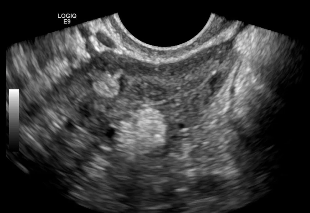 Ultrasound
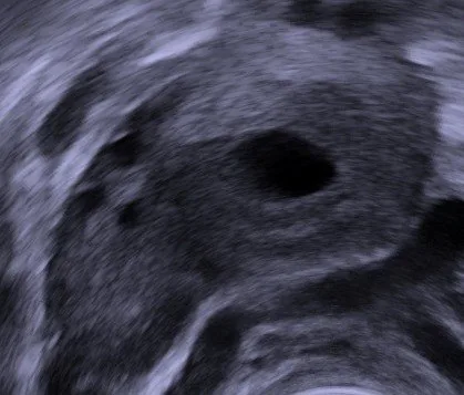 Ultrasound
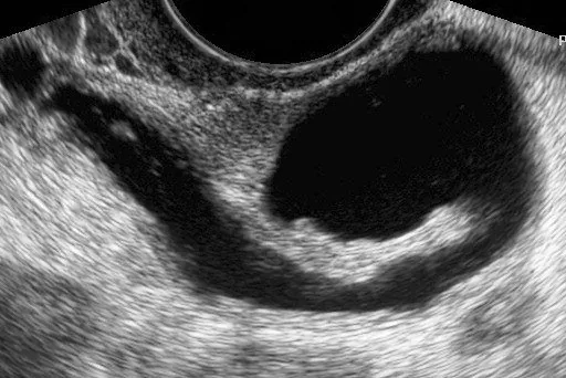 Ultrasound
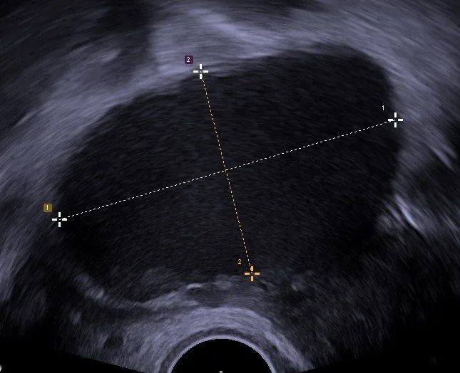 Ultrasound
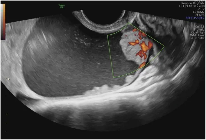 Ultrasound
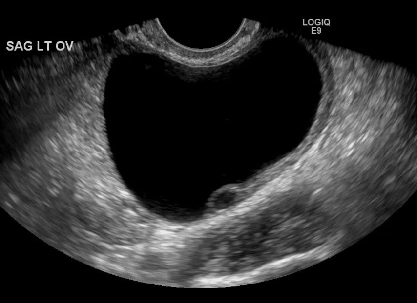 Ultrasound
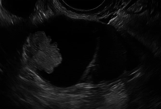 Ultrasound
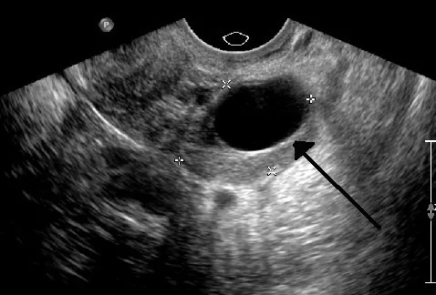 Ultrasound
Classification
Functional Cysts
- The follicular cyst is unilocular, anechoic, smooth-walled, and usually resolves within two to three cycles.
- The corpus luteum shows a thick, highly vascularised wall with a 'ring of fire' on Doppler and an anechoic or haemorrhagic centre.
- The haemorrhagic cyst characteristically has a fine reticular fishnet pattern or a retracted clot without its own blood flow.
Dermoid and Endometrioma
- The mature teratoma is identified by the echogenic Rokitansky nodule, fat-fluid levels, and fine echogenic lines and dots from hair.
- The endometrioma appears on ultrasound as homogeneous and hypoechoic with ground-glass internal echoes and no blood flow in the internal components.
- Dermoids are particularly prone to torsion because their contents are dense and heavy.
- CA 125 is also elevated in endometriosis, which limits its diagnostic value in young women.
Diagnosis
Non-Ovarian Origin
- A hydrosalpinx shows a convoluted, anechoic tubular structure with incomplete septation and the 'cogwheel sign'.
- A tubo-ovarian abscess is a complex cystic-solid mass with fever, signs of inflammation, and cervical motion tenderness.
- A peritoneal inclusion cyst is identified by the fact that the ovary lies freely within the fluid and the wall is formed by the peritoneum.
What IOTA is
- IOTA acronym: The acronym IOTA stands for International Ovarian Tumour Analysis, a collaborative group for the standardised description of adnexal findings.
- Described features: The IOTA standard always describes size in three planes, internal structure, septa, solid components, papillary projections, ascites and vascularisation.
Easy Descriptors
- B-descriptor: A typical B-descriptor is, for example, the unilocular cyst with a smooth wall in a premenopausal woman.
- M-descriptor: A typical M-descriptor is, for example, ascites plus a solid tumour with vascularisation in a postmenopausal woman.
Simple Rules
- Structure: The Simple Rules consist of five benign and five malignant features that are weighed against each other.
- Benign features (B-rules): The benign features include unilocular cyst, solid component < 7 mm, acoustic shadows, smooth-walled multilocular tumour < 10 cm and no blood flow.
- Malignant features (M-rules): The malignant features include irregular solid tumour, ascites, at least four papillary projections, irregular multilocular-solid tumour > 10 cm and very strong blood flow.
Typical imaging findings
- Ultrasound: A roughly five-centimetre, completely anechoic structure with a smooth thin wall lies within the ovary. The tissue behind it is markedly brighter (posterior acoustic enhancement). No septa, no solid components, no papillary projections.
- Ultrasound: A round, anechoic structure with a smooth, thin wall, no solid components and no relevant internal echoes — the typical appearance of a simple (functional) ovarian cyst.
Keep learning in the app
Further reading (selection)
Cross-references
More topics: Ovary & adnexa
- Corpus luteum cyst and haemorrhagic ovarian cyst
- Mature cystic teratoma (dermoid cyst)
- Adnexal torsion (ovarian torsion)
- Ovarian cancer
- Borderline ovarian tumour
- Ovarian germ cell tumours
- Granulosa cell tumour and sex cord-stromal tumours
- Krukenberg tumour
- Pelvic inflammatory disease (PID)
- Tubo-ovarian abscess
Note: Learning content from the GynFuchs app (flashcards, exam questions, image cases) for medical education – not a treatment recommendation and no substitute for diagnosis or treatment decisions in individual cases. Treatment and management are deliberately not covered on this page.