Meigs syndrome
Exam relevance: in 2 of 197 board exam reports · rank 144
- Specialty
- Gynaecology · Ovary & adnexa
- Images
- Mammography/MRI 1 · Endoscopy & gross 1 · Histology & cytology 1
- Exam relevance
- 2 of 197 reports · rank 144
- Last updated
- 10/2026 · Dr. Pascal Bafteh
Contents
Images (3)
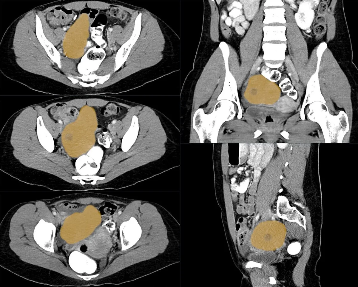 Mammography/MRI
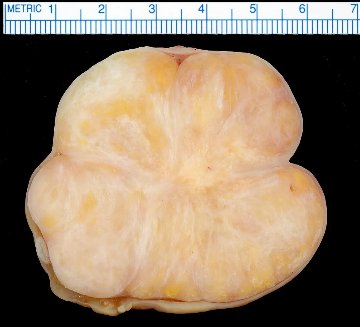 Endoscopy & gross
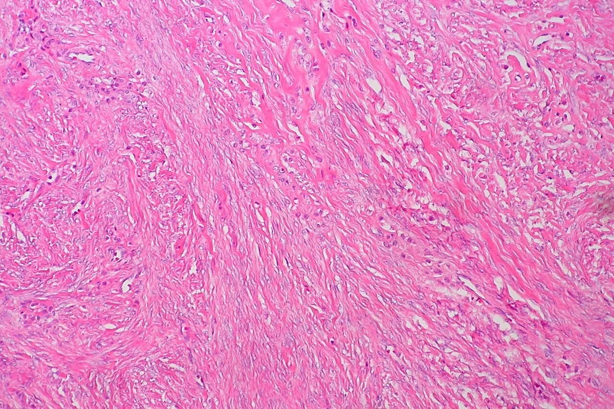 Histology & cytology
Definition
- Meigs syndrome denotes a benign ovarian tumour, classically a fibroma, with ascites and pleural effusion.
- The criteria established by Meigs in 1954 comprise a benign ovarian tumour such as fibroma, thecoma, granulosa cell tumour or Brenner tumour, ascites, pleural effusion and regression of both effusions once the tumour is no longer present.
Classification
- If another tumour is the cause, such as other benign ovarian tumours like struma ovarii, mucinous cystadenoma or teratoma, a uterine leiomyoma or ovarian metastases, the term pseudo-Meigs syndrome is used.
Occurrence & epidemiology
Epidemiology and pathogenesis
- The syndrome affects about 1% of ovarian tumours; ascites is found in 10–15% and hydrothorax in 1% of women with ovarian fibroma.
- It is very uncommon before the third decade and increases thereafter to a peak in the seventh decade; most patients are postmenopausal.
- The exact pathogenesis is unknown; it is thought that fluid passes from the peritoneal cavity through diaphragmatic defects or lymphatic channels into the pleural space, causing an exudative effusion.
Clinical features
- Typical are dyspnoea, dry cough and painful abdominal distension; the pleural effusion is right-sided in about 70%, left-sided or bilateral in 15% each.
Diagnosis
- Examination reveals an adnexal mass, diminished breath sounds and ascites; imaging confirms the pelvic mass.
- Ultrasound shows ascites, possibly pleural effusions and a well-demarcated adnexal mass without increased vascularity.
- CA 125 may be raised; the mechanism is unclear, mesothelial expression is suspected.
- The definitive diagnosis rests on resolution of the effusions and histological confirmation of the tumour.
Keep learning in the app
Further reading (selection)
Cross-references
More topics: Ovary & adnexa
- Ovarian cysts (adnexal cysts)
- Corpus luteum cyst and haemorrhagic ovarian cyst
- Mature cystic teratoma (dermoid cyst)
- Adnexal torsion (ovarian torsion)
- Ovarian cancer
- Borderline ovarian tumour
- Ovarian germ cell tumours
- Granulosa cell tumour and sex cord-stromal tumours
- Krukenberg tumour
- Pelvic inflammatory disease (PID)
Note: Learning content from the GynFuchs app (flashcards, exam questions, image cases) for medical education – not a treatment recommendation and no substitute for diagnosis or treatment decisions in individual cases. Treatment and management are deliberately not covered on this page.