Mature cystic teratoma (dermoid cyst)
Exam relevance: in 9 of 197 board exam reports · rank 75
- Specialty
- Gynaecology · Ovary & adnexa
- Images
- Clinical 1 · Ultrasound 1 · Endoscopy & gross 1
- Exam relevance
- 9 of 197 reports · rank 75
- Last updated
- 10/2026 · Dr. Pascal Bafteh
Contents
Images (3)
Definition
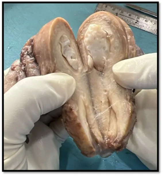
- Mature cystic teratoma (dermoid cyst) is a benign germ cell tumour composed of tissues from all three germ layers; ectodermal tissue and sebaceous material are present in almost every case.
Classification
- Besides the mature cystic teratoma as the commonest form, there are monodermal teratomas such as struma ovarii and carcinoid tumours, and fetiform teratomas.
Occurrence & epidemiology
Epidemiology
- It is the most common ovarian germ cell tumour in young women; teratomas account for about 20% of all ovarian neoplasms.
- Patients are younger than those with epithelial tumours, with a mean age of about 30 years; bilateral involvement is reported in 8–15%, and growth is about 1.8 mm per year.
Clinical features
Clinical features and complications
- The tumour is often detected incidentally on imaging or pelvic examination; chronic pelvic pain or a palpable mass may occur, and acute pain indicates a complication.
- Complications are torsion (16%), rupture (1–4%) with possible chemical peritonitis, malignant transformation (0.5–3%), infection and, rarely, autoimmune haemolytic anaemia or anti-NMDA receptor encephalitis; malignant change mainly affects older women.
Histology
- The cyst wall bears squamous epithelium; the cavity is filled with sebaceous material that is liquid at body temperature.
Diagnosis
Typical imaging findings
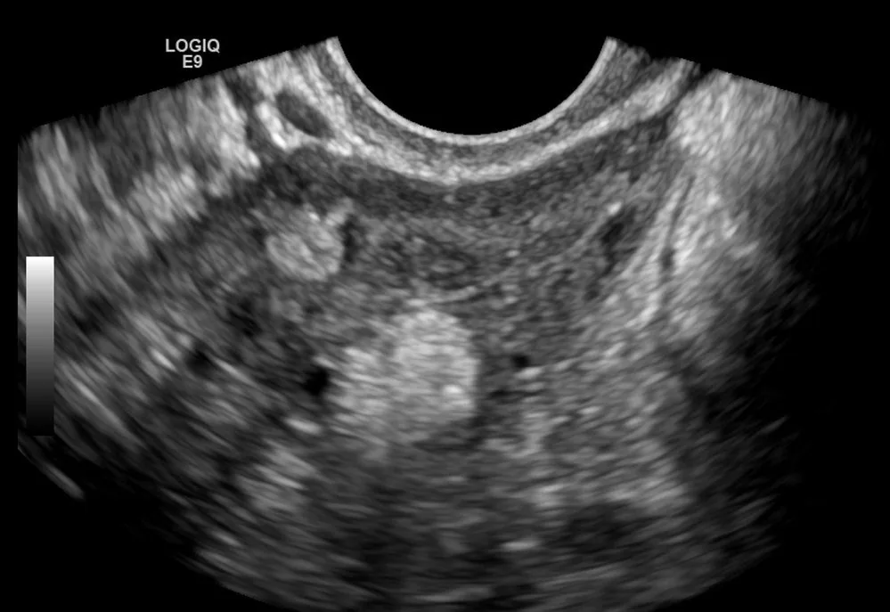
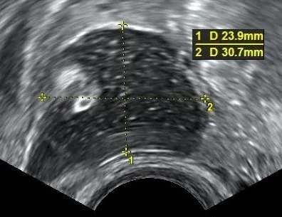
- Ultrasound: An adnexal mass of mixed echogenicity is seen. A strongly echogenic, rounded nodule stands out from the rest of the contents and casts an acoustic shadow; beside it are fine echogenic lines and dots floating within the cyst.
- Ultrasound: A round, predominantly hyperechoic mass with a heterogeneous internal structure on the ovary — consistent with a mature cystic teratoma (dermoid cyst) containing fatty components.
- On ultrasound, the dermoid plug (Rokitansky nodule), the dot-dash pattern from hair, the tip of the iceberg sign and fat-fluid levels are typical.
- On CT and MRI the detection of fat is diagnostic; on MRI the fatty layer is T1-hyperintense and loses signal on fat-suppressed sequences.
Keep learning in the app
Further reading (selection)
Cross-references
More topics: Ovary & adnexa
Note: Learning content from the GynFuchs app (flashcards, exam questions, image cases) for medical education – not a treatment recommendation and no substitute for diagnosis or treatment decisions in individual cases. Treatment and management are deliberately not covered on this page.