Peritoneal carcinomatosis of gynaecological origin
Exam relevance: in 12 of 197 board exam reports · rank 59
- Specialty
- Gynaecology · Ovary & adnexa
- Images
- Endoscopy & gross 2 · Mammography/MRI 1 · Histology & cytology 2
- Exam relevance
- 12 of 197 reports · rank 59
- Last updated
- 10/2026 · Dr. Pascal Bafteh
Contents
Images (5)
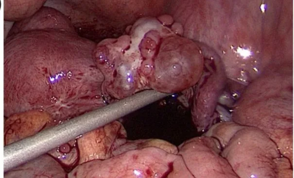 Endoscopy & gross
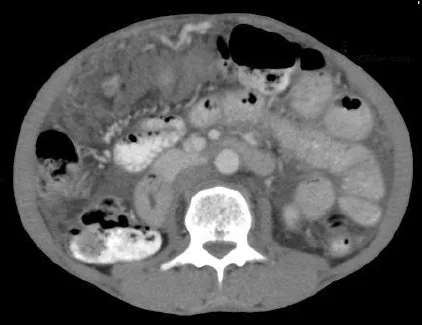 Mammography/MRI
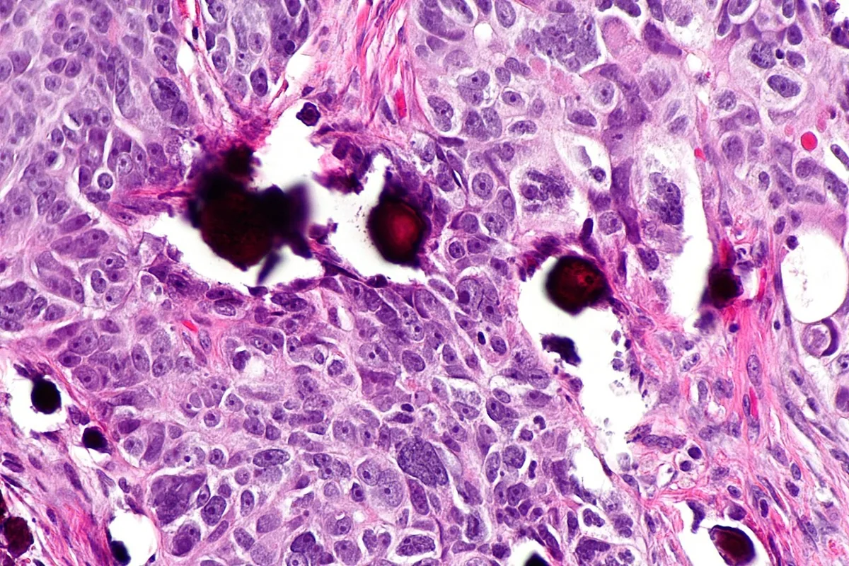 Histology & cytology
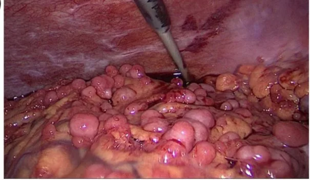 Endoscopy & gross
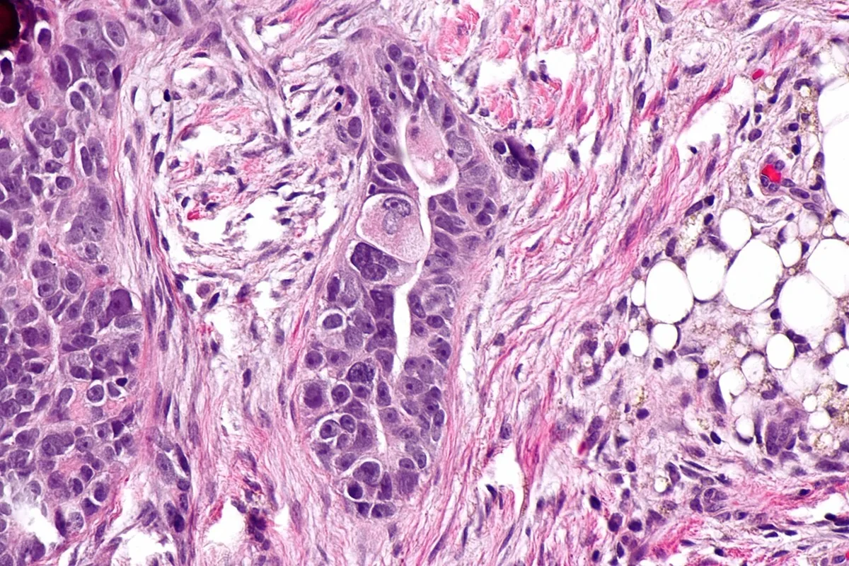 Histology & cytology
Definition
- Peritoneal surface malignancies are tumours arising from or spreading to the peritoneum, including primary peritoneal tumours, pseudomyxoma peritonei and peritoneal carcinomatosis from gastrointestinal or gynaecological primaries.
- Peritoneal carcinomatosis is the implantation of neoplastic cells in the peritoneal cavity; it is often multifocal, appearing as widespread nodules or mucinous deposits.
Classification
- Mucinous, non-mucinous and primary peritoneal forms are distinguished.
- The Sugarbaker Peritoneal Cancer Index (PCI) records the distribution and size of deposits in nine abdominopelvic regions and four small bowel regions.
- In each region the lesion size is graded: no tumour, nodules under 0.5 cm, 0.5–5 cm or over 5 cm.
Occurrence & epidemiology
Epidemiology
- In ovarian cancer about 70% of patients are diagnosed at an advanced stage (FIGO III–IV) or with peritoneal carcinomatosis.
- Peritoneal carcinomatosis is also relatively frequent in advanced stages of many gastrointestinal tumours such as colonic, gastric and pancreatic cancer.
Aetiopathogenesis
Pathogenesis
- Ovarian cancer usually spreads within the abdomen via the peritoneal circulation.
- Peritoneal fluid flows from the pelvis via the paracolic gutters to the subdiaphragmatic regions, drawn by negative pressure as the diaphragm rises during expiration; on the right it reaches the anterior subhepatic and right hepatic spaces.
- On the left the phrenicocolic ligament limits flow towards the perisplenic spaces.
Clinical features
- Complications are malignant ascites, bowel obstruction and malnutrition.
Diagnosis
- According to international guidelines CT is the standard imaging modality for ovarian cancer staging, with an accuracy of up to 94%, and assesses the primary tumour, peritoneal implants, lymph nodes and distant metastases.
- MRI helps where CT is inadequate, such as subphrenic spaces, the lesser omentum and serosal and mesenteric deposits.
- The diagnosis relies on cross-sectional imaging, histology of the implants and tumour markers such as CEA or CA 125; laparoscopy can confirm the extent and allow tissue sampling.
Keep learning in the app
Further reading (selection)
Cross-references
More topics: Ovary & adnexa
- Ovarian cysts (adnexal cysts)
- Corpus luteum cyst and haemorrhagic ovarian cyst
- Mature cystic teratoma (dermoid cyst)
- Adnexal torsion (ovarian torsion)
- Ovarian cancer
- Borderline ovarian tumour
- Ovarian germ cell tumours
- Granulosa cell tumour and sex cord-stromal tumours
- Krukenberg tumour
- Pelvic inflammatory disease (PID)
Note: Learning content from the GynFuchs app (flashcards, exam questions, image cases) for medical education – not a treatment recommendation and no substitute for diagnosis or treatment decisions in individual cases. Treatment and management are deliberately not covered on this page.