Breast cancer
Exam relevance: in 77 of 197 board exam reports · rank 1
- Synonyms
- breast carcinoma
- Specialty
- Gynaecology · Breast
- Images
- Ultrasound 4 · Mammography/MRI 4 · Endoscopy & gross 2 · Clinical 2
- Exam relevance
- 77 of 197 reports · rank 1
- In the app
- 5 flashcards · GynFuchs
- Last updated
- 10/2026 · Dr. Pascal Bafteh
Contents
Images (12)
 Ultrasound
 Mammography/MRI
 Endoscopy & gross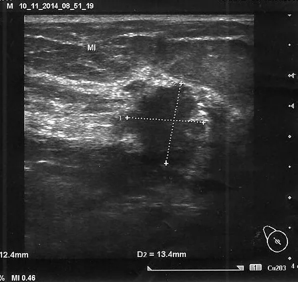
 Ultrasound
 Mammography/MRI
 Endoscopy & gross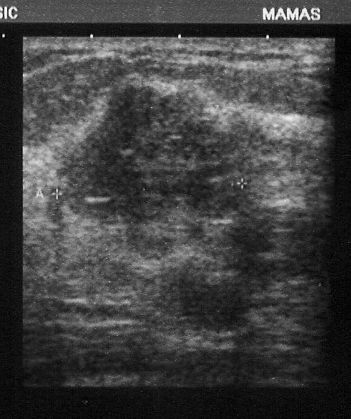
 Ultrasound
 Mammography/MRI
 Ultrasound
 Mammography/MRI
Classification
Molecular Subtypes
- Luminal A: A Luminal A carcinoma is hormone receptor-positive, HER2-negative, well-differentiated with a low Ki-67.
- Luminal B: A Luminal B carcinoma is hormone receptor-positive with a high Ki-67 or poorer grade, HER2 negative or positive.
- Triple-negative: In triple-negative breast cancer: oestrogen receptor, progesterone receptor, and HER2 are all negative.
Aetiopathogenesis
BRCA and HRD
- BRCA1/2 function: BRCA1 and BRCA2 are tumour suppressor genes involved in homologous recombination, which repair DNA double-strand breaks.
- HRD definition: HRD means homologous recombination deficiency, i.e., the functional defect of this repair pathway even without a BRCA mutation.
- Germline vs. somatic mutation: A germline mutation differs from a somatic mutation in that it is present in all body cells and can be inherited.
Clinical features
History
- Regarding the finding itself, questions are asked about duration, change in size, pain, cyclical nature, and nipple discharge.
- The family history records breast and ovarian cancers in relatives and their respective age at diagnosis.
- The age of affected relatives at diagnosis is important because it is part of the score for indicating BRCA testing.
Inspection
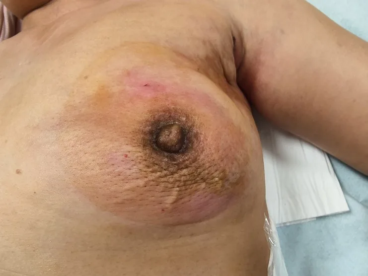
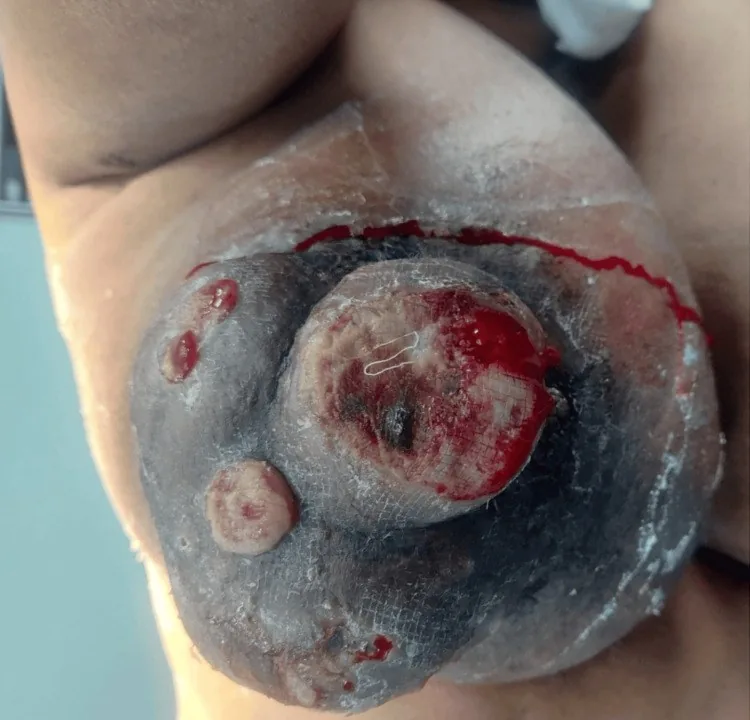
- In a sitting position, with arms lowered and raised, one looks for asymmetry, bulging, plateau sign, skin dimpling, and nipple retraction.
- Peau d'orange is caused by lymphatic congestion in the cutis, which tethers the skin at the hair follicles.
Palpation
- Technically, one palpates flat with the finger pads against a counter-pressure, systematically in all quadrants, in sitting and supine positions.
- A lump typical of carcinoma is firm, poorly defined, has limited mobility, and is mostly painless.
- Also palpated are the axilla as well as the infraclavicular and supraclavicular regions bilaterally.
- The most common location is the upper outer quadrant, accounting for about half of all carcinomas.
More facts from the study questions
- The typical palpation findings of a breast carcinoma are a firm consistency, poor demarcation from surrounding tissue and low mobility.
- The correct sequence is: mammography before core needle biopsy.
- A haematoma caused by the biopsy can significantly impair the interpretability of a subsequent mammogram.
- After a suspicious palpable finding, ultrasound of both breasts and the axillae is the first imaging step.
- It serves to further characterise the finding.
- Approximately half of all breast carcinomas are located in the upper outer quadrant.
- This area is examined with particular care during palpation.
- An eczematous, scaly, erosive change of the nipple is suspicious for Paget's disease of the nipple.
- The Elston and Ellis grading system is an established system for assessing the aggressiveness of breast carcinoma.
- It is based on the histological evaluation of three criteria: tubule formation, nuclear pleomorphism and mitotic rate.
Histology
Mandatory Information
- Histological type: The pathology report first states the histological type, i.e. invasive, no special type (NST), invasive lobular, or a special type.
- Grading according to Elston and Ellis: This assesses the three criteria of tubule formation, nuclear pleomorphism, and mitotic rate.
- Hormone receptor status: This includes the oestrogen and progesterone receptor status as a percentage of stained tumour cell nuclei.
- HER2 status: The HER2 status is determined by immunohistochemistry and re-tested by FISH if 2+.
- Ki-67: The proliferation marker Ki-67 measures the proportion of proliferating cells and thus the growth rate.
Diagnosis
Five malignant signs
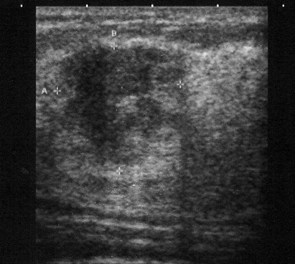
- Shape: A carcinoma is typically irregular, angular or spiculated.
- Orientation: The orientation of a malignant lesion to the skin is mostly vertical, i.e., taller-than-wide.
- Internal echoes: The internal echoes of a carcinoma are characteristically markedly hypoechoic and inhomogeneous.
- Posterior phenomenon: Posterior to the lesion, there is often an acoustic shadow because the tumour is sound-absorbing and desmoplastic.
- Surroundings: The surroundings of a carcinoma show an echogenic halo and infiltration instead of displacement.
Axilla and Doppler
- Normal lymph node: An unremarkable axillary lymph node has a broad, echogenic fatty hilum and a narrow, hypoechoic cortex.
- Lymph node suspicious for malignancy: A lymph node is suspicious for malignancy if it is round, shows an effaced hilum and an eccentrically thickened cortex.
- Colour Doppler: In carcinoma, Colour Doppler often shows prominent, irregularly penetrating peripheral vessels, but is not conclusive on its own.
Limitations of Ultrasound
- Necessity of mammography: Mammography remains indispensable because ultrasound does not reliably detect microcalcifications and thus DCIS.
- Difficult to detect on mammography: Often difficult to detect on mammography are lobular carcinomas and LCIS, because they grow diffusely and show little density difference.
What the abbreviations stand for
- BI-RADS stands for Breast Imaging Reporting and Data System of the American College of Radiology.
- ACR in this context stands for the American College of Radiology, which defines density categories a to d.
Categories
- BI-RADS 0 means assessment incomplete, further imaging or prior images are needed.
- BI-RADS 4 means suspicious, subdivided into 4a to 4c — histological clarification required.
Density
- ACR a and b describe predominantly fatty involuted glandular tissue with good mammographic assessability.
- ACR c and d describe dense glandular tissue where the sensitivity of mammography is reduced.
- In practice, high density means a higher risk of interval cancer and the importance of supplementary ultrasound.
Typical imaging findings
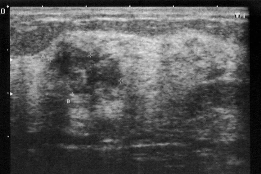
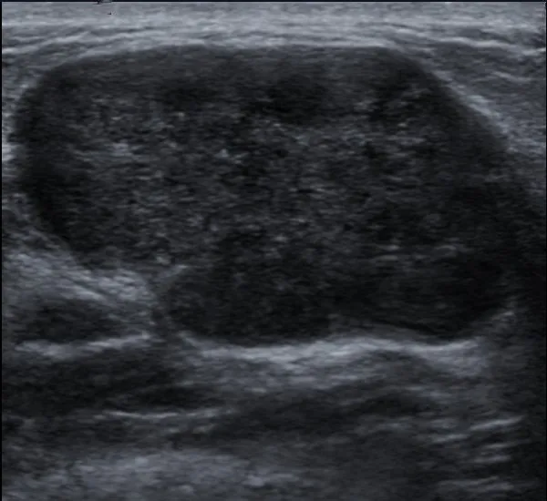
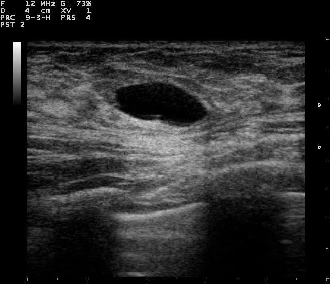
- Ultrasound: The hypoechoic lesion stands perpendicular to the skin and is larger in depth than in width. Its margins are ill-defined and the internal structure heterogeneous; the calipers show the vertical diameter to be the larger one.
- Ultrasound: The lesion is hypoechoic with ill-defined, angular margins and a heterogeneous internal structure. Marked posterior shadowing is seen behind it; the surrounding tissue planes appear interrupted and calipers span the lesion.
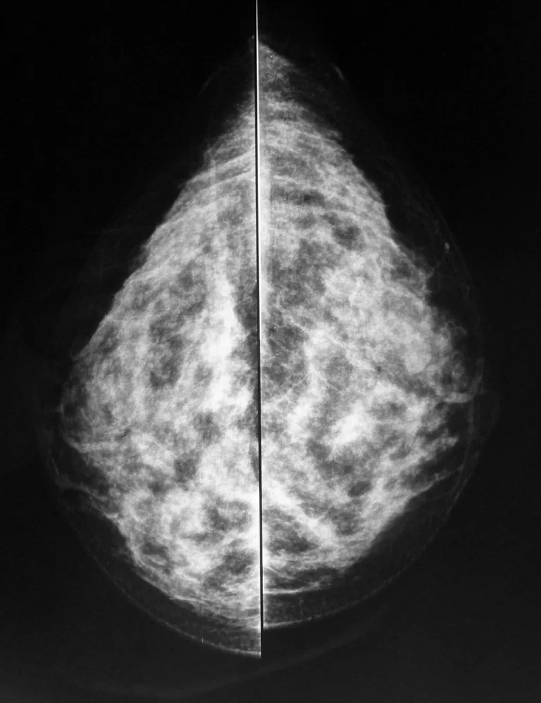
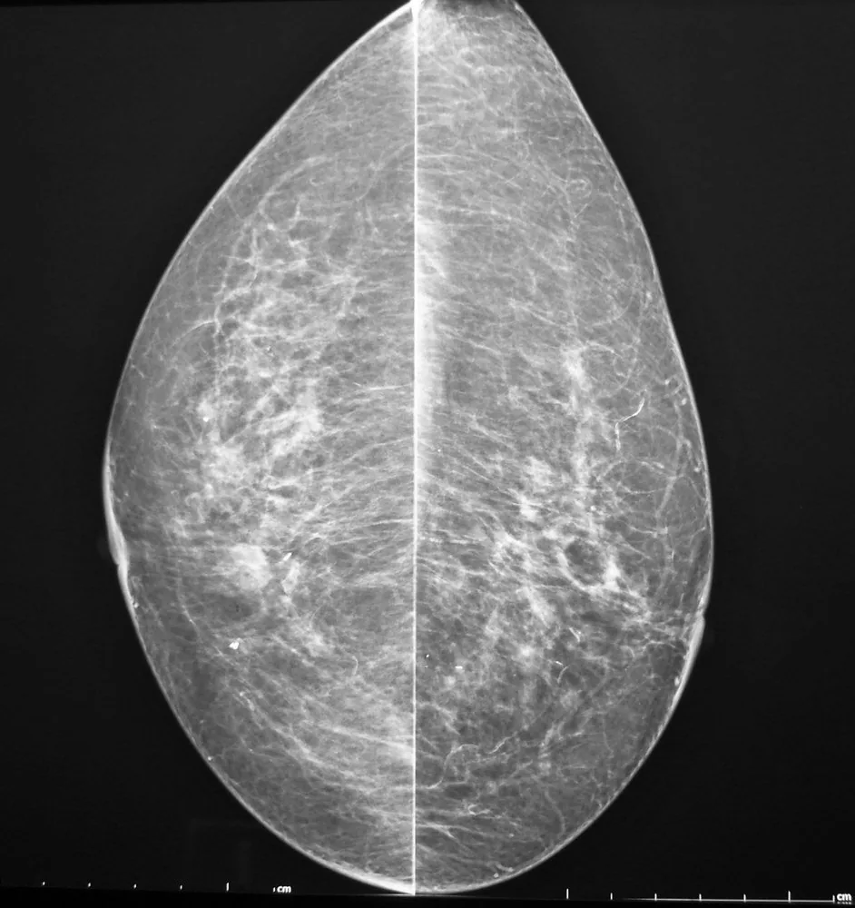
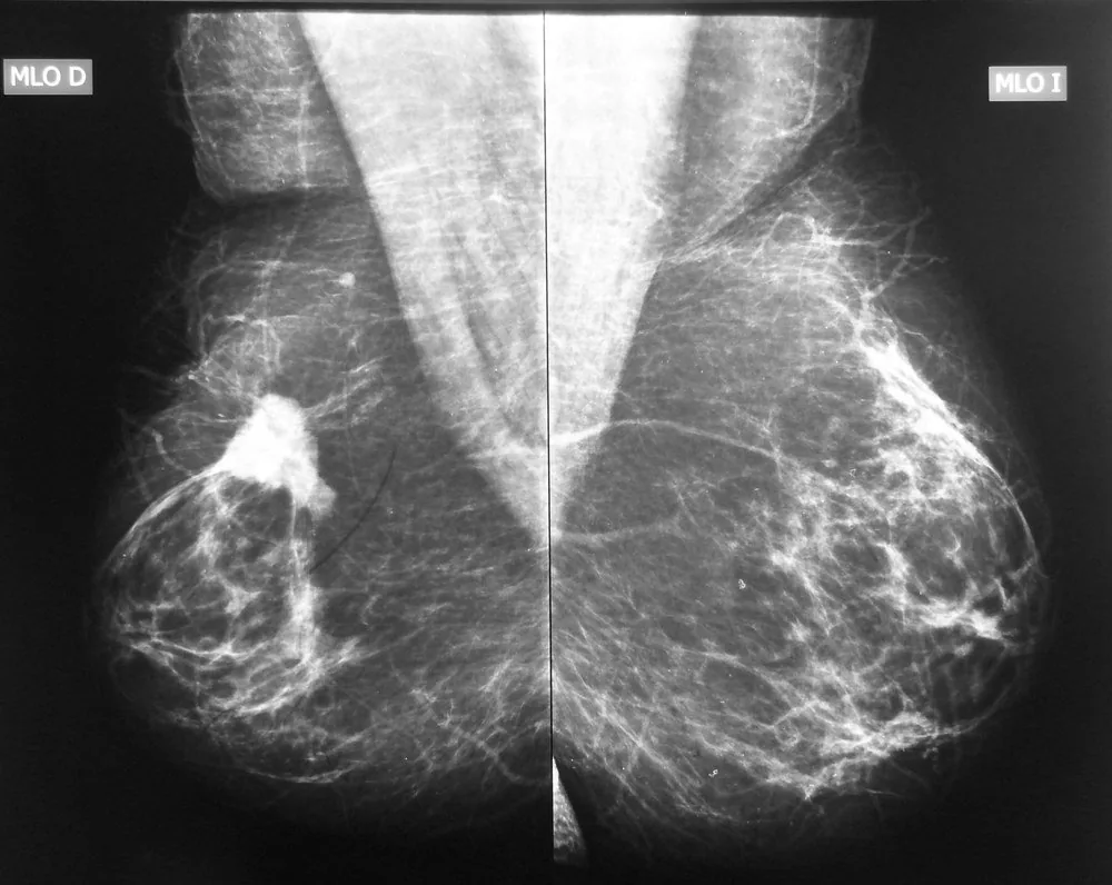
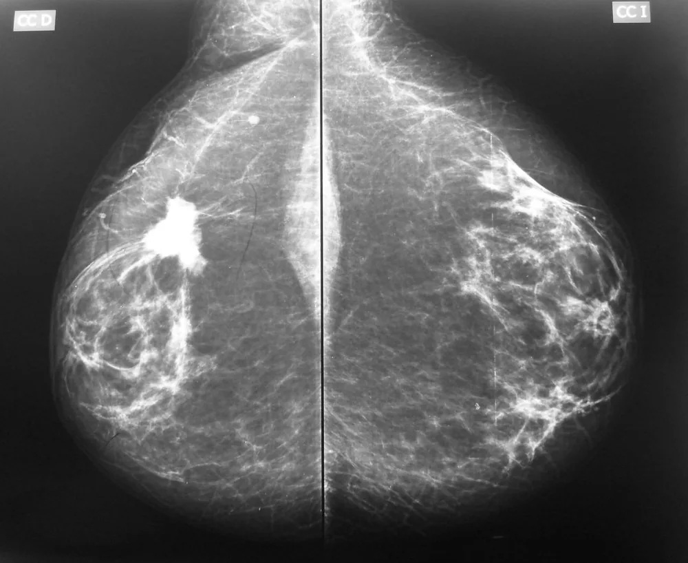
- Mammography: Fine strands of differing length radiate in a stellate pattern from a dense centre into the surrounding tissue. The normal connective tissue strands are pulled towards this point; the lesion has no smooth margin and no bright halo.
- Mammography: On the right there is a dense, irregularly bordered mass with fine spicules radiating into the surrounding tissue in a stellate fashion. The left side is normal by comparison; no microcalcification is visible within the lesion.
Keep learning in the app
Further reading (selection)
Cross-references
More topics: Breast
- Triple-negative breast cancer
- HER2-positive breast cancer
- Lobular breast cancer and lobular neoplasia
- B3 lesions of the breast
- Hereditary breast and ovarian cancer syndrome (BRCA)
- Ductal carcinoma in situ (DCIS)
- Fibroadenoma
- Fibrocystic breast changes and breast cysts
- Intraductal papilloma
- Non-puerperal mastitis
Note: Learning content from the GynFuchs app (flashcards, exam questions, image cases) for medical education – not a treatment recommendation and no substitute for diagnosis or treatment decisions in individual cases. Treatment and management are deliberately not covered on this page.