Fibroadenoma
Exam relevance: in 12 of 197 board exam reports · rank 59
- Synonyms
- breast lump
- Specialty
- Gynaecology · Breast
- Images
- Clinical 4 · Ultrasound 4
- Exam relevance
- 12 of 197 reports · rank 59
- In the app
- 2 flashcards · GynFuchs
- Last updated
- 10/2026 · Dr. Pascal Bafteh
Contents
Images (8)
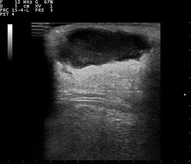
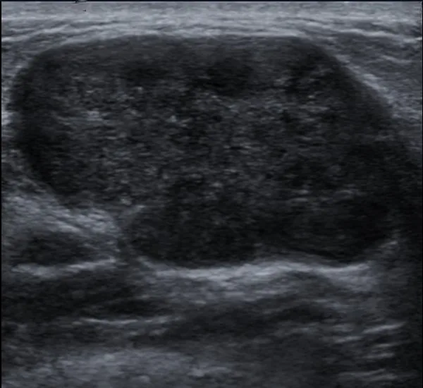 Ultrasound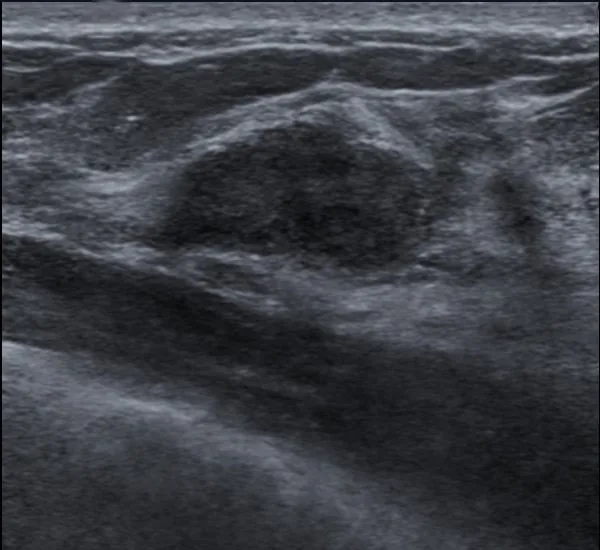
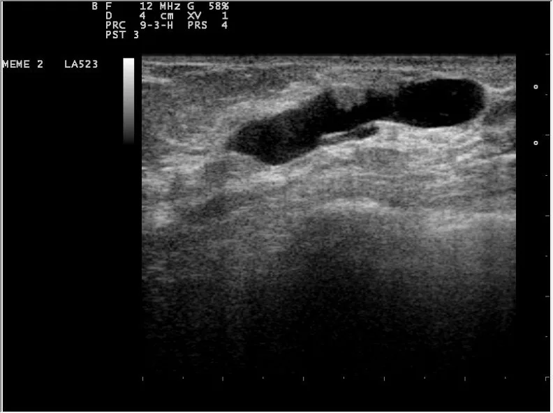 Ultrasound
 Ultrasound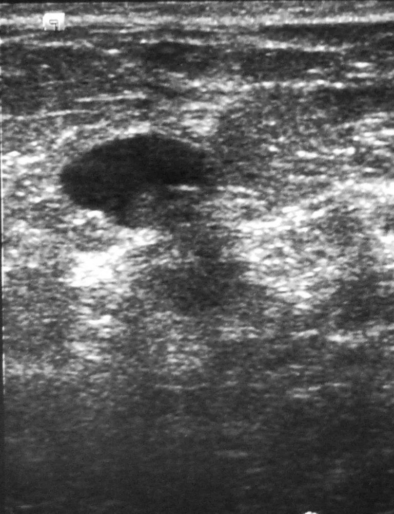
 Ultrasound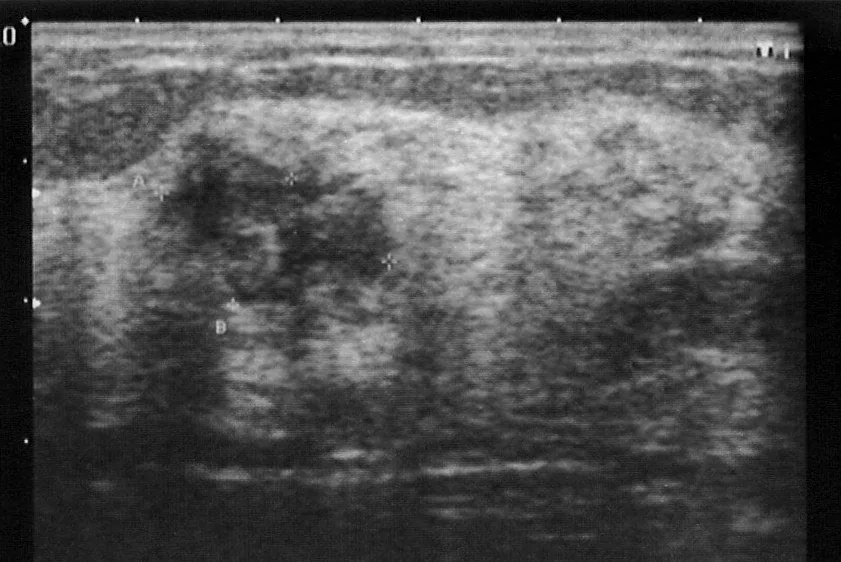
Definition
Lumps in the breast
- A fibroadenoma is the most common benign breast tumour in young women, firm, well-circumscribed, and mobile.
- Benign, solid breast tumour composed of epithelial and stromal tissue.
Occurrence & epidemiology
Epidemiology
- Fibroadenoma is the most common benign breast tumour.
- It occurs most commonly between 14 and 35 years but can be found at any age; it is less common after the menopause.
Aetiopathogenesis
Aetiology
- Fibroadenomas are hormone dependent: they enlarge with increased oestrogen and progesterone activity, in pregnancy and lactation, and decrease in size around the menopause.
Clinical features
- Typical is a painless, unilateral, rubbery, smoothly bordered and highly mobile lump (breast mouse).
Histology
- Histology shows proliferation of ductal epithelium and stroma with intracanalicular and pericanalicular growth patterns.
- A fibroadenoma is termed complex when cysts larger than 3 mm, sclerosing adenosis, epithelial calcifications or papillary apocrine metaplasia are present; it tends to occur in older women.
Diagnosis
Benign features on ultrasound
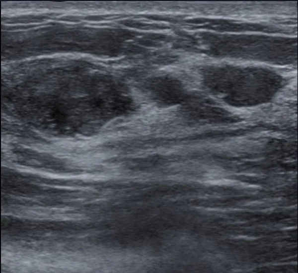
- Fibroadenoma: A typical fibroadenoma is oval, oriented parallel to the skin, with smooth or gently lobulated margins and homogeneously hypoechoic.
- Cyst: A simple cyst is sonographically anechoic, well-defined and shows posterior acoustic enhancement with lateral edge shadows.
- Growth pattern: A displacing rather than infiltrating growth pattern suggests benignity.
Typical imaging findings
- Ultrasound: The lesion is oval, smoothly and gently lobulated, and clearly wider than tall, that is parallel to the skin. Its internal structure is homogeneously hypoechoic; there is mild posterior enhancement and no shadowing.
- Ultrasound typically shows a smooth, oval, hypoechoic mass.
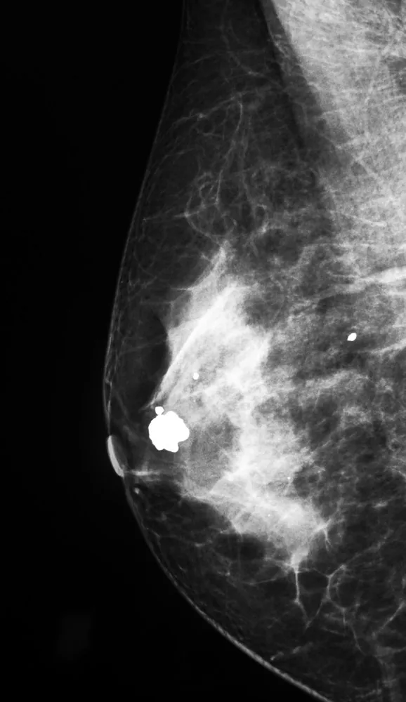
- Densely calcified fibroadenomas show the typical coarse popcorn pattern on mammography and CT.
Keep learning in the app
Further reading (selection)
- StatPearls: Breast Fibroadenoma (NCBI Bookshelf)
- Cytomorphological Features of Complex Fibroadenoma Breast (J Cytol 2023, PubMed Central)
- Beyond the lung: recognizing incidental breast lesions on chest computed tomography (Transl Cancer Res 2026, PubMed Central)
- DocCheck Flexikon, Mastitis non-puerperalis
- DocCheck Flexikon, Mammasonographie
Cross-references
More topics: Breast
- Breast cancer
- Triple-negative breast cancer
- HER2-positive breast cancer
- Lobular breast cancer and lobular neoplasia
- B3 lesions of the breast
- Hereditary breast and ovarian cancer syndrome (BRCA)
- Ductal carcinoma in situ (DCIS)
- Fibrocystic breast changes and breast cysts
- Intraductal papilloma
- Non-puerperal mastitis
Note: Learning content from the GynFuchs app (flashcards, exam questions, image cases) for medical education – not a treatment recommendation and no substitute for diagnosis or treatment decisions in individual cases. Treatment and management are deliberately not covered on this page.