Paget disease of the nipple
- Specialty
- Gynaecology · Breast
- Images
- Clinical 1
- Last updated
- 10/2026 · Dr. Pascal Bafteh
Contents
Images (1)
Definition
- Eczema-like change of the nipple and areola caused by large intraepidermal carcinoma cells (Paget cells), associated in over 90% with underlying DCIS or invasive breast cancer.
Occurrence & epidemiology
Epidemiology
- About 1–3% of breast cancers present in this way; mainly postmenopausal women aged 50 to 60 are affected, and rarely men.
Aetiopathogenesis
Aetiology and pathogenesis
- According to the most widely accepted epidermotropic theory, Paget cells migrate from an underlying carcinoma along the lactiferous ducts into the epidermis of the nipple.
- The transformation theory instead regards Paget cells as malignantly transformed keratinocytes of the nipple epidermis.
Clinical features
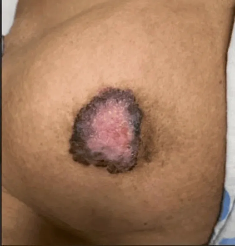
- Typical is a well-defined, irregularly shaped, red, scaly or crusted patch that starts on the nipple and spreads to the areola; usually only one side is affected.
- Pain, itching and burning occur in 15–25% and often precede the visible changes; ulceration, bleeding and discharge tend to occur in advanced stages.
- A palpable mass may be present and indicates a high probability of invasive cancer.
Histology
- Paget cells are large cells with abundant pale cytoplasm, pleomorphic nuclei and prominent nucleoli and lie singly or in clusters within the epidermis.
- On immunohistochemistry they are typically positive for low-molecular-weight cytokeratins such as CK7 and CAM5.2; HER2 is overexpressed in 80–90%, and oestrogen and progesterone receptors are positive in about 40% and 30%, respectively.
Diagnosis
Typical imaging findings
- Clinical finding: The nipple itself is destroyed and replaced by a sharply demarcated, eroded, reddish, scaly surface that was confirmed on biopsy to show Paget cells.
- Clinical finding: The nipple itself is replaced by a sharply demarcated, red, eroded surface that extends onto the areola. The surface is crusted and the nipple body partly destroyed.
- The diagnosis is confirmed by a full-thickness biopsy of the nipple-areola complex with histology and immunohistochemistry, while mammography, ultrasound and MRI look for a lesion within the breast.
- Invasive cancer may be present even when imaging is unremarkable.
Keep learning in the app
Further reading (selection)
Cross-references
More topics: Breast
Note: Learning content from the GynFuchs app (flashcards, exam questions, image cases) for medical education – not a treatment recommendation and no substitute for diagnosis or treatment decisions in individual cases. Treatment and management are deliberately not covered on this page.