Fibrocystic breast changes and breast cysts
Exam relevance: in 2 of 197 board exam reports · rank 144
- Synonyms
- fibrocystic breast, breast cyst
- Specialty
- Gynaecology · Breast
- Images
- Ultrasound 1 · Mammography/MRI 2 · Clinical 2
- Exam relevance
- 2 of 197 reports · rank 144
- Last updated
- 10/2026 · Dr. Pascal Bafteh
Contents
Images (5)
 Ultrasound
 Mammography/MRI
 Mammography/MRI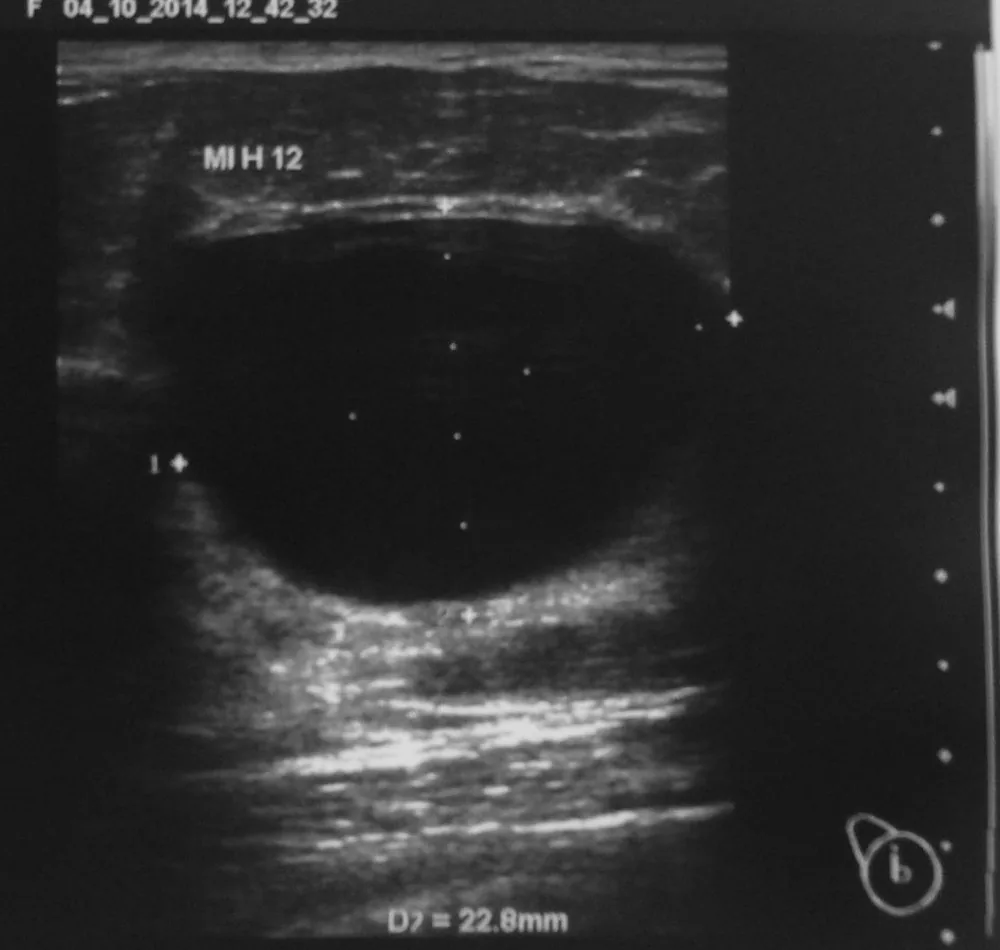
Definition
- The most common benign breast condition, now usually termed fibrocystic changes rather than disease; it reflects hormonally mediated remodelling of breast tissue and is not a single disease entity.
Occurrence & epidemiology
Epidemiology
- Fibrocystic changes affect a substantial proportion of women, particularly during the reproductive years, and cause the most commonly reported breast symptoms; symptoms tend to subside after menopause.
Aetiopathogenesis
Aetiology and pathogenesis
- Cyclical fluctuations of oestrogen and progesterone during the reproductive years influence epithelial proliferation, stromal oedema and cyst formation.
- Repeated stimulation by oestrogen and progesterone probably contributes; the changes are more common with early menarche, nulliparity or a first live birth after age 30.
Clinical features
- The breasts have a nodular, dense texture, are frequently tender and feel heavy; some women describe a burning pain.
- Cysts and nondescript masses usually lie in the upper outer part of the breast; painful, tender, rubbery masses in women of reproductive age with a history of similar findings suggest fibrocystic changes.
Histology
- Nonproliferative changes include simple cysts, duct ectasia, apocrine metaplasia, mild epithelial hyperplasia without proliferation and stromal fibrosis.
- Nonproliferative changes are distinguished from proliferative lesions without and with atypia; nonproliferative findings do not raise breast cancer risk significantly, proliferative lesions without atypia raise it about 1.5- to 2-fold and atypical hyperplasia about 4- to 5-fold.
Diagnosis
Typical imaging findings
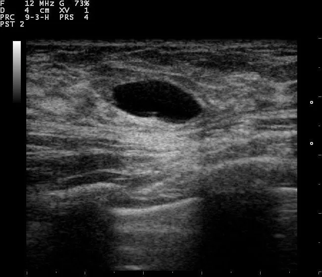
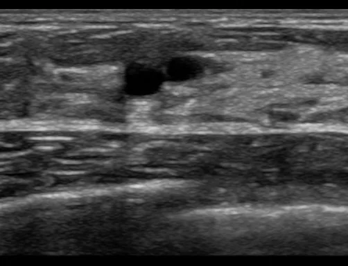
- Ultrasound: The lesion is completely anechoic, very sharply and smoothly bordered, with marked posterior acoustic enhancement. Narrow lateral shadows are seen; internal echoes, septa or solid components are absent.
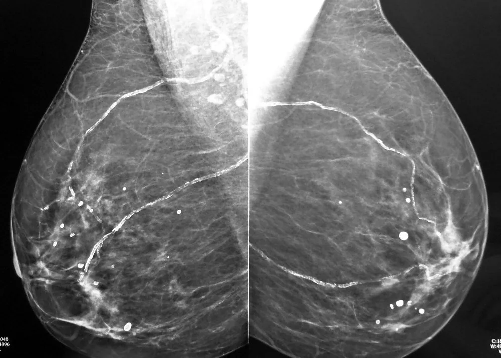
- Mammography: Two parallel fine lines of calcification run like tram tracks over long distances through both breasts; beside them lie individual round, smoothly bordered calcifications. No clustered fine calcification, no mass.
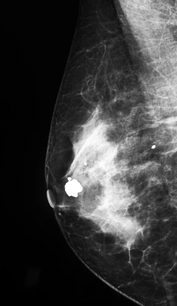
- Mammography: Within a smoothly bordered mass lies a coarse, densely calcified structure reminiscent of popcorn. The calcification is large and irregular in shape but sharply demarcated; the surrounding tissue is normal.
- Ultrasound is the preferred initial imaging modality in younger women and dense breast tissue and distinguishes cystic from solid lesions; mammography serves to exclude cancer.
- Solid masses are confirmed histologically by image-guided core biopsy.
Keep learning in the app
Further reading (selection)
Cross-references
Note: Learning content from the GynFuchs app (flashcards, exam questions, image cases) for medical education – not a treatment recommendation and no substitute for diagnosis or treatment decisions in individual cases. Treatment and management are deliberately not covered on this page.