Ductal carcinoma in situ (DCIS)
Exam relevance: in 10 of 197 board exam reports · rank 66
- Specialty
- Gynaecology · Breast
- Images
- Mammography/MRI 3 · Clinical 4
- Exam relevance
- 10 of 197 reports · rank 66
- In the app
- 1 flashcards · GynFuchs
- Last updated
- 10/2026 · Dr. Pascal Bafteh
Contents
Images (7)
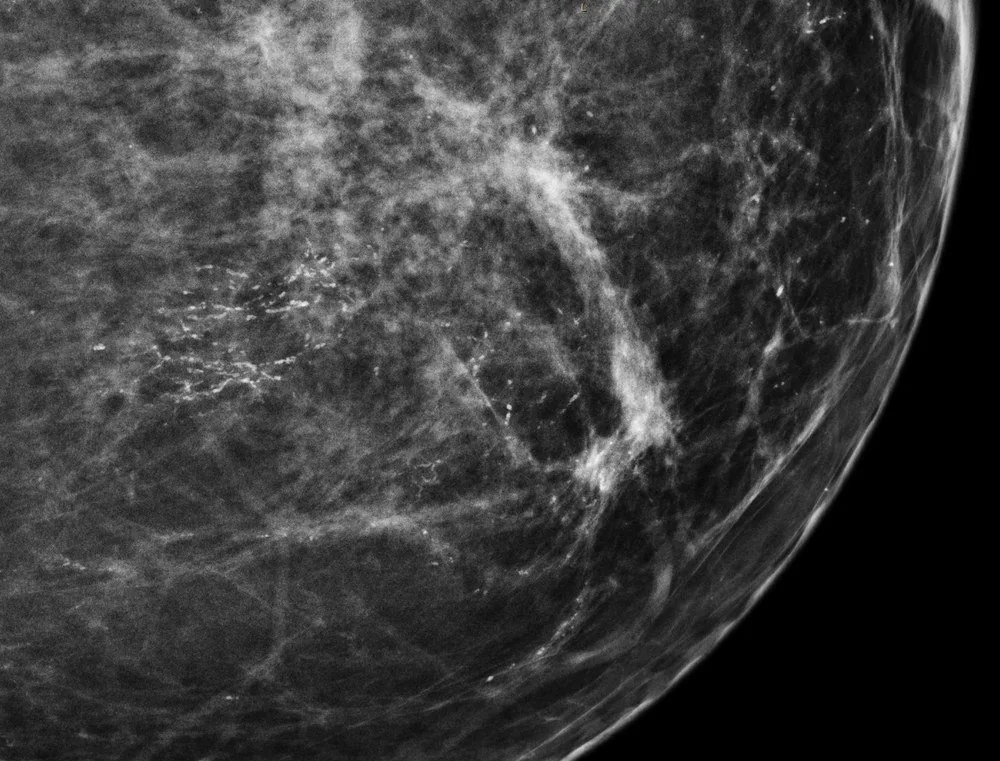 Mammography/MRI
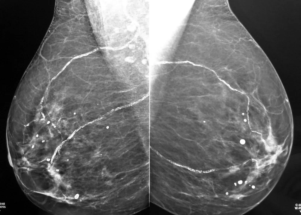 Mammography/MRI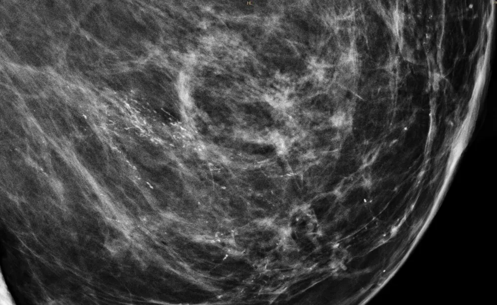
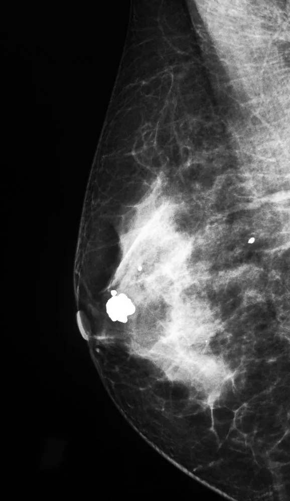 Mammography/MRI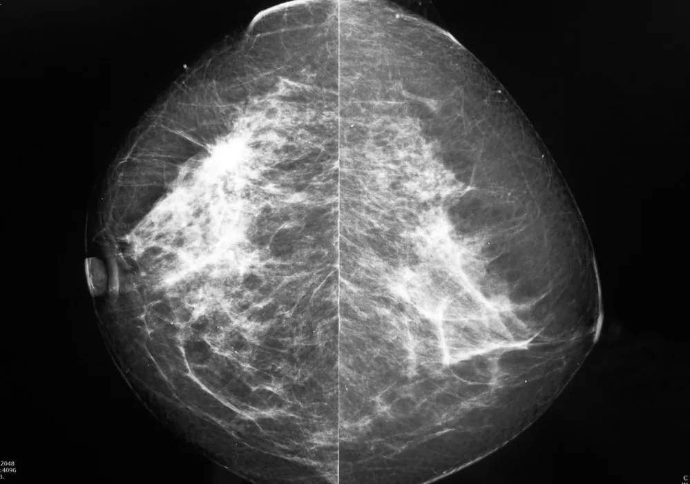
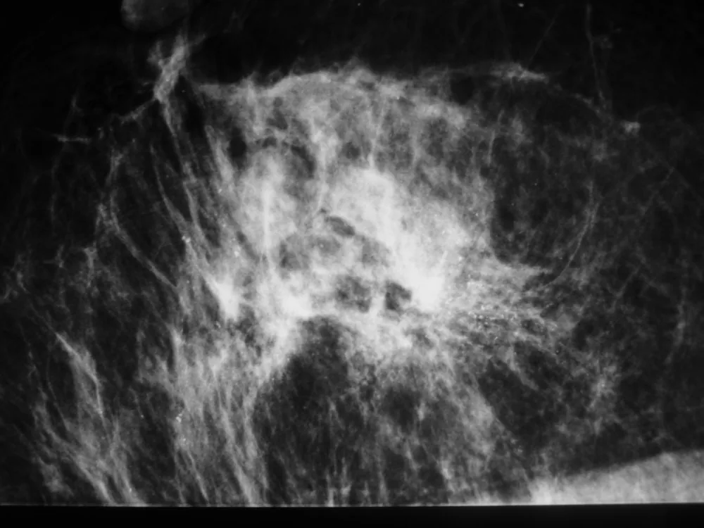
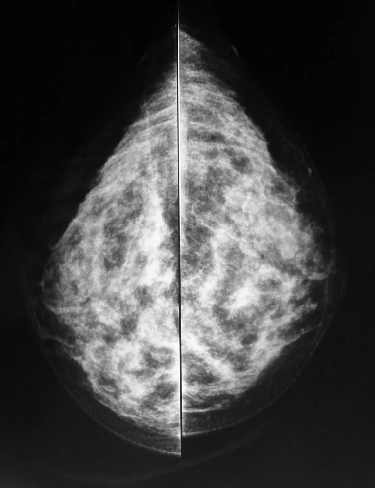
Definition
What DCIS is
- Definition: Ductal carcinoma in situ (DCIS) is an intraductal neoplasia in which the basement membrane is intact, thus making metastasis impossible.
- Radiological appearance: On mammography, DCIS almost always presents as clustered, pleomorphic microcalcifications in a ductal or segmental distribution.
- Sonographic appearance: On ultrasound, DCIS is usually not clearly identifiable — which is precisely why screening requires mammography.
Diagnosis
Benign vs. Malignant Calcification
- Criteria for malignancy: Features suggestive of malignancy are fine, pleomorphic or linear-branching calcifications in a clustered, segmental arrangement.
- Criteria for benignity: Considered benign are coarse 'popcorn' calcifications of a fibroadenoma, vascular calcifications with a 'tram-track' appearance, 'eggshell' and 'milk of calcium' calcifications.
- Further assessment: For further assessment of suspicious calcifications, magnification and compression views of the calcified area are used.
Work-up
- Biopsy: In cases of suspicious microcalcifications, the standard procedure is stereotactically guided vacuum-assisted biopsy.
Typical imaging findings
- Mammography: Within a circumscribed area numerous very fine calcific particles of differing size and shape lie densely together. They are arranged along an imaginary line towards the nipple; there is no palpable or visible mass.
Keep learning in the app
Further reading (selection)
Cross-references
More topics: Breast
Note: Learning content from the GynFuchs app (flashcards, exam questions, image cases) for medical education – not a treatment recommendation and no substitute for diagnosis or treatment decisions in individual cases. Treatment and management are deliberately not covered on this page.