B3 lesions of the breast
Exam relevance: in 4 of 197 board exam reports · rank 111
- Specialty
- Gynaecology · Breast
- Images
- Ultrasound 2
- Exam relevance
- 4 of 197 reports · rank 111
- In the app
- 1 flashcards · GynFuchs
- Last updated
- 10/2026 · Dr. Pascal Bafteh
Contents
Images (2)
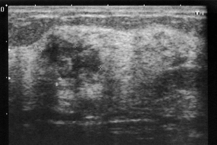 Ultrasound
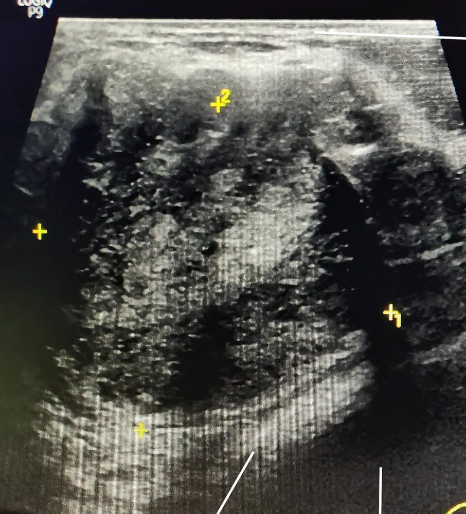 Ultrasound
Definition
- B3 lesions are breast lesions of uncertain malignant potential: a heterogeneous group with a borderline histological spectrum and a variable but low risk of associated malignancy.
Classification
B classification
- B classification: The classification from B1 to B5 describes the assessment of malignancy potential of the core biopsy material, from normal tissue to malignancy.
- B3 lesions: B3 lesions with uncertain malignant potential include atypical ductal hyperplasia, flat epithelial atypia, lobular neoplasia, and papilloma.
- They include atypical ductal hyperplasia (ADH), flat epithelial atypia, classical lobular neoplasia, papillary lesions, benign phyllodes tumours and radial scars.
- If carcinoma is found in subsequent tissue, DCIS is categorised as B5a and invasive carcinoma as B5b.
Occurrence & epidemiology
Epidemiology
- Between 4 and 9% of all core needle biopsies are classified as B3, with numbers increasing due to MRI and vacuum-assisted biopsy; the positive predictive value for malignancy has fallen from 29 to 10%.
- Atypical hyperplasia (ADH and atypical lobular hyperplasia) is found in 4–10% of benign breast biopsies performed for imaging findings or palpable lesions.
Clinical features
- ADH is usually found incidentally on needle biopsy specimens obtained because of abnormal mammographic findings; flat epithelial atypia mostly appears as microcalcifications on mammography.
Histology
- Histologically ADH essentially resembles low-grade DCIS and is distinguished by extent: below 2 mm it is diagnosed as ADH, above 2 mm as low-grade DCIS.
- ADH increases breast cancer risk about fivefold; it is regarded as a risk lesion rather than a precursor lesion, because the associated carcinoma can arise anywhere in either breast and not only at the site of the ADH.
Diagnosis
- The diagnosis is made on core needle biopsy (mostly 14G) or vacuum-assisted biopsy (7G–11G) under ultrasound, stereotactic or MRI guidance.
- The rate of carcinoma in subsequent tissue varies by lesion; in the consensus conference data it was highest for ADH (27.6%) and considerably lower for papillary lesions (7.8%).
Keep learning in the app
Further reading (selection)
Cross-references
More topics: Breast
Note: Learning content from the GynFuchs app (flashcards, exam questions, image cases) for medical education – not a treatment recommendation and no substitute for diagnosis or treatment decisions in individual cases. Treatment and management are deliberately not covered on this page.